You are currently on the International (English) version.
Looks like you are in United States.
Switch to local content?
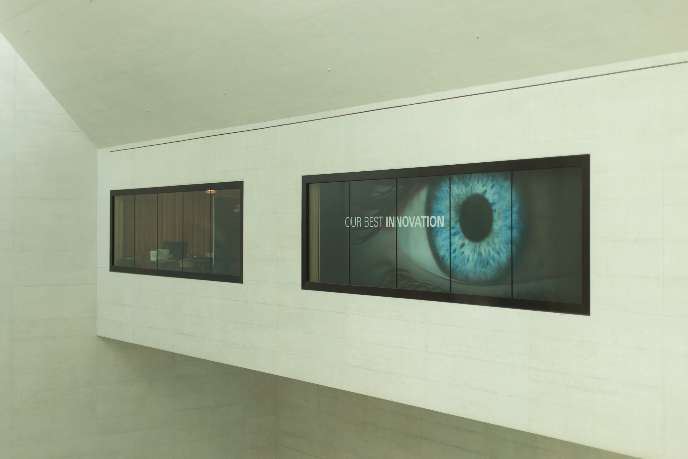
Heidelberg Engineering joins the Collaborative Community on Ophthalmic Innovation (CCOI) through EssilorLuxottica’s Vision Architect membership
Heidelberg Engineering today announced that it is joining the Collaborative Community on Ophthalmic Innovation (CCOI) as part of its parent company EssilorLuxottica’s Vision Architect membership.
All News & Stories
Where cutting-edge ophthalmic technology meets real-world clinical impact.

Heidelberg Engineering Appoints Senior Medical Director Ophthalmology to Further Strengthen the Company’s Commitment to Innovation

Heidelberg Engineering Highlights Innovation, Collaboration, and Research at ARVO 2026
Heidelberg Engineering announces expansion of market access for digital surgical visualization platform

Advancing AI in Retinal Imaging: Hrvoje Bogunović Receives Xtreme Research Award

Heidelberg Engineering UK Announces Restructure to Capitalise on Future Growth

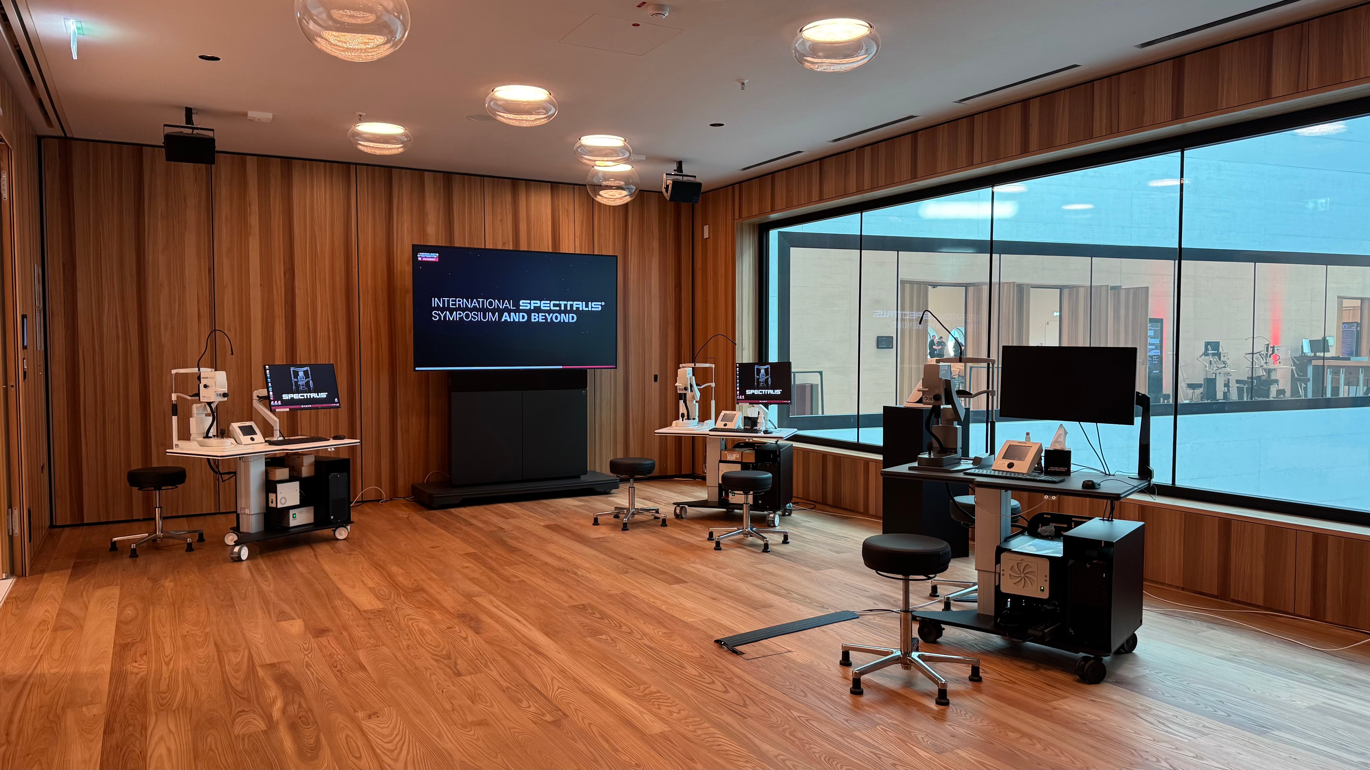
International Scientific Symposium 2026 Marks a New Chapter in Ophthalmic Imaging

Heidelberg Engineering to Establish Direct Subsidiary in Spain with Appointment of José Manuel Tamarit

Heidelberg Engineering’s Tosh Vadhia Announced as New Ambassador for Orbis UK
Heidelberg Engineering Reflects on a Year of Milestones and Looks Ahead to 2026
Heidelberg Engineering joins the Collaborative Community on Ophthalmic Innovation (CCOI) through EssilorLuxottica’s Vision Architect membership


Global Experts Unite: Visionary Horizons Sets the Stage for the Future of Ophthalmology
| 2 Minutes
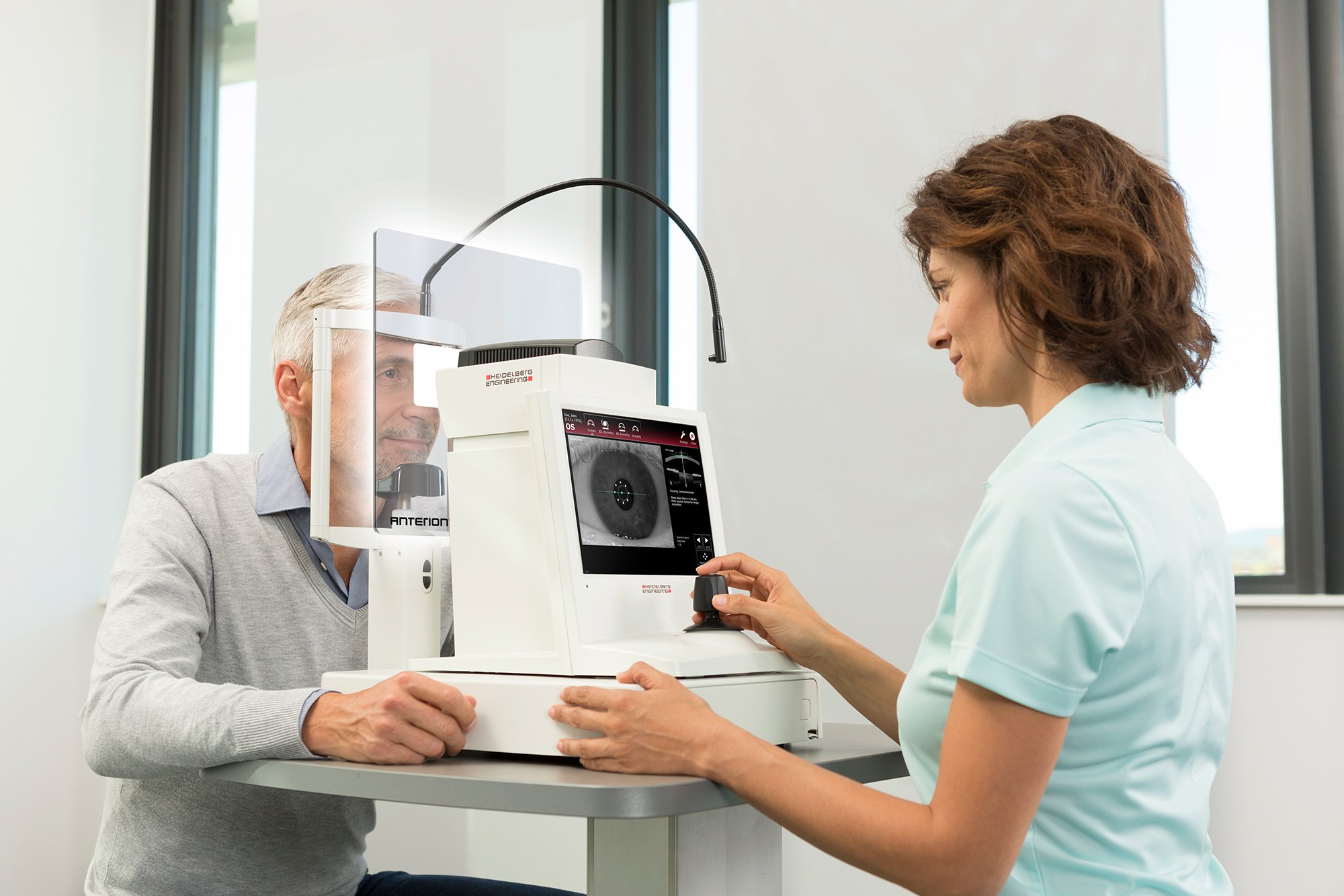
Grow Your Practice Through Anterior Segment Specialization
Heidelberg Engineering at ESCRS 2025: New ANTERION® Feature Streamlines Cataract Workflow and Fosters Ecosystem Integration
Taking your OCT Outside of the Posterior Pole
Heidelberg Engineering expands Leadership Team with Appointments of Dr. Stéphanie Magazzeni and Dr. Holger Ruchatz

Orbis and Heidelberg Engineering Announce Expansion of Collaboration During a Special Visit to the Flying Eye Hospital in Rwanda
Henrik Martinsson Leads Heidelberg Engineering’s Direct Presence in Sweden

Heidelberg Engineering Celebrates Eye2Gene™ AI Breakthrough in Precision Ophthalmology
Insights from ARVO 2025