You are currently on the United Kingdom version.
Looks like you are in United States.
Switch to local content?
Connecting Cataract, Cornea, and Glaucoma Care in One Platform
From cataract and refractive surgery to corneal and glaucoma diagnostics, ANTERION® integrates precise OCT imaging and measurements into smooth, efficient clinical workflows.
One Platform for Many Stages of Anterior Segment Care
ANTERION® supports every stage of anterior segment care: From diagnostics to surgical planning and follow-up. With precise OCT imaging, reproducible measurements, and intuitive visualisation, it adapts seamlessly to the clinical workflow of each specialty.
Cataract Surgery –
Confident Planning From the Start
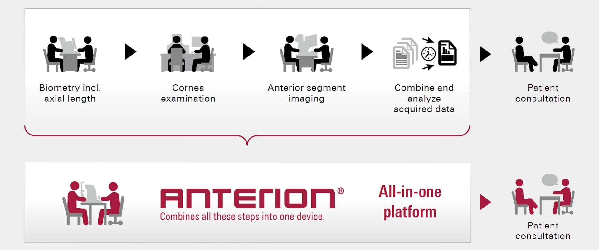
ANTERION® provides all key measurements for refractive cataract surgery planning in one patient examination. Precise biometry and detailed cornea as well as anterior chamber analysis support accurate IOL calculation and lens selection. Visual confirmation of each parameter adds confidence, even in challenging or post-refractive eyes. With integrated IOL calculators and seamless data export, ANTERION® simplifies the pre-operative workflow and helps achieve predictable refractive outcomes.
Refractive Surgery –
Precise Corneal Data for Individualised Planning
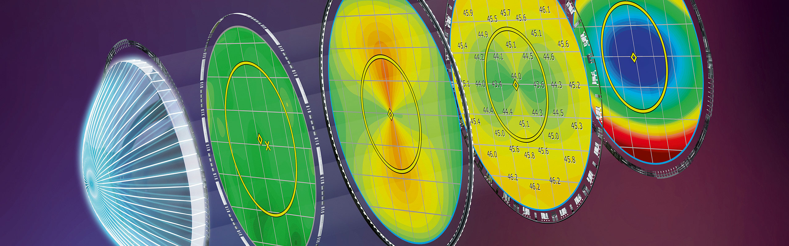
For refractive surgeons, ANTERION® delivers a complete OCT-based corneal analysis that supports every step of the refractive planning workflow. High-resolution tomography of the anterior and posterior surfaces, pachymetry, and wavefront data provide a full understanding of corneal shape and thickness distribution. Epithelial thickness mapping and difference maps help identify irregularities, assess stability, and guide personalised treatment planning for safe, predictable refractive outcomes. The integrated Ectasia View and the SCORE Analyser assist in evaluating corneal integrity and assessing ectasia risk before and after surgery.
Cornea and Ectasia Management –
Comprehensive Corneal Insight
For cornea specialists, ANTERION® offers a complete OCT-based analysis of anterior and posterior surfaces to assess full corneal geometry. Detailed curvature, elevation, pachymetry, and epithelial thickness maps enable precise assessment of alterations, keratoconus and post-surgical changes. Tools such as Ectasia View and the SCORE Analyser* help identify risk and monitor progression over time. Customisable layouts and follow-up comparisons streamline decision-making and documentation throughout the corneal diagnostic workflow.
*Powered by Dr. Gatinel and Dr. Saad for AInsight
Glaucoma and Anterior Chamber Assessment – Detailed Angle Evaluation
ANTERION® visualises the entire anterior chamber in high-resolution OCT and automatically detects the scleral spur for precise angle metrics. Quantitative parameters such as AOD, TISA, and ACA support non-contact assessment of angle-closure risk and surgical planning. The 360° angle view and refraction-corrected measurements provide reproducible data for longitudinal follow-up, complementing gonioscopy and enhancing diagnostic confidence in glaucoma management.