You are currently on the United Kingdom version.

Looks like you are in United States.

Switch to local content?

Apps Optimised to the Clinical Need

ANTERION® adapts to your clinical needs with different applications. Each ANTERION® App builds on high-resolution OCT imaging to deliver precise data, advanced analysis options, and workflow efficiency across anterior segment diagnostics.

Customise Your ANTERION® with These Apps

Built for versatility, ANTERION® offers modular Apps that expand your clinical capability. From imaging, biometry and cornea tomography to anterior chamber analysis, each App provides precise data and smooth integration into your workflow.

Cataract App

All essential biometry parameters in one acquisition. The Cataract App combines corneal analysis, anterior chamber depth, lens thickness, and axial length with IOL power calculation tools.

1 / 5

Biometry and IOL Power Calculation for Refractive Cataract Surgery Planning

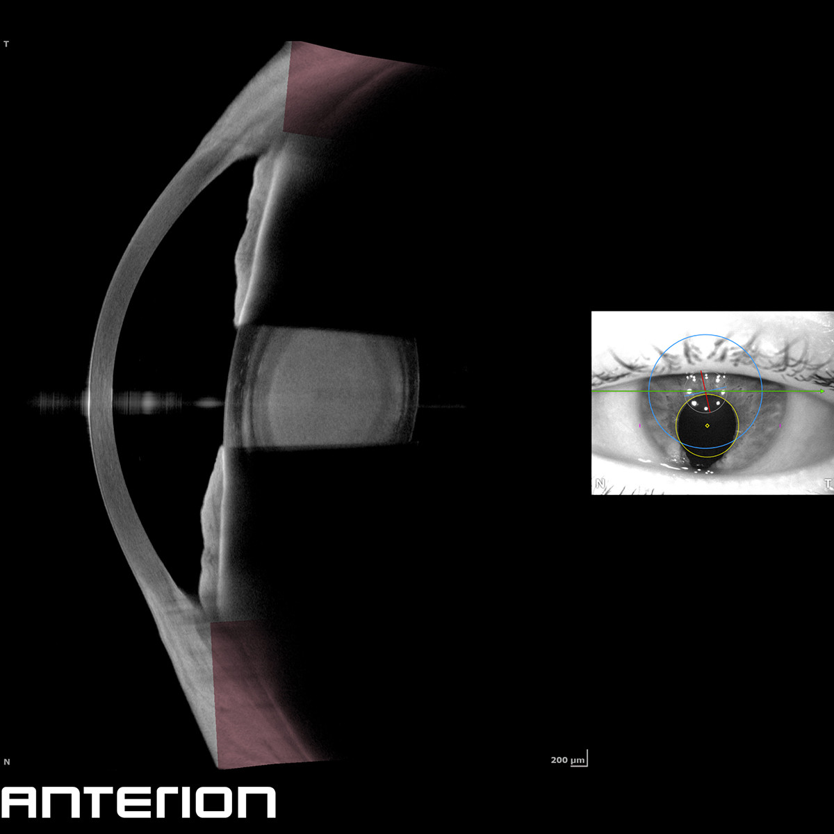
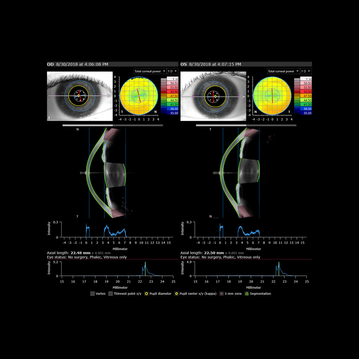
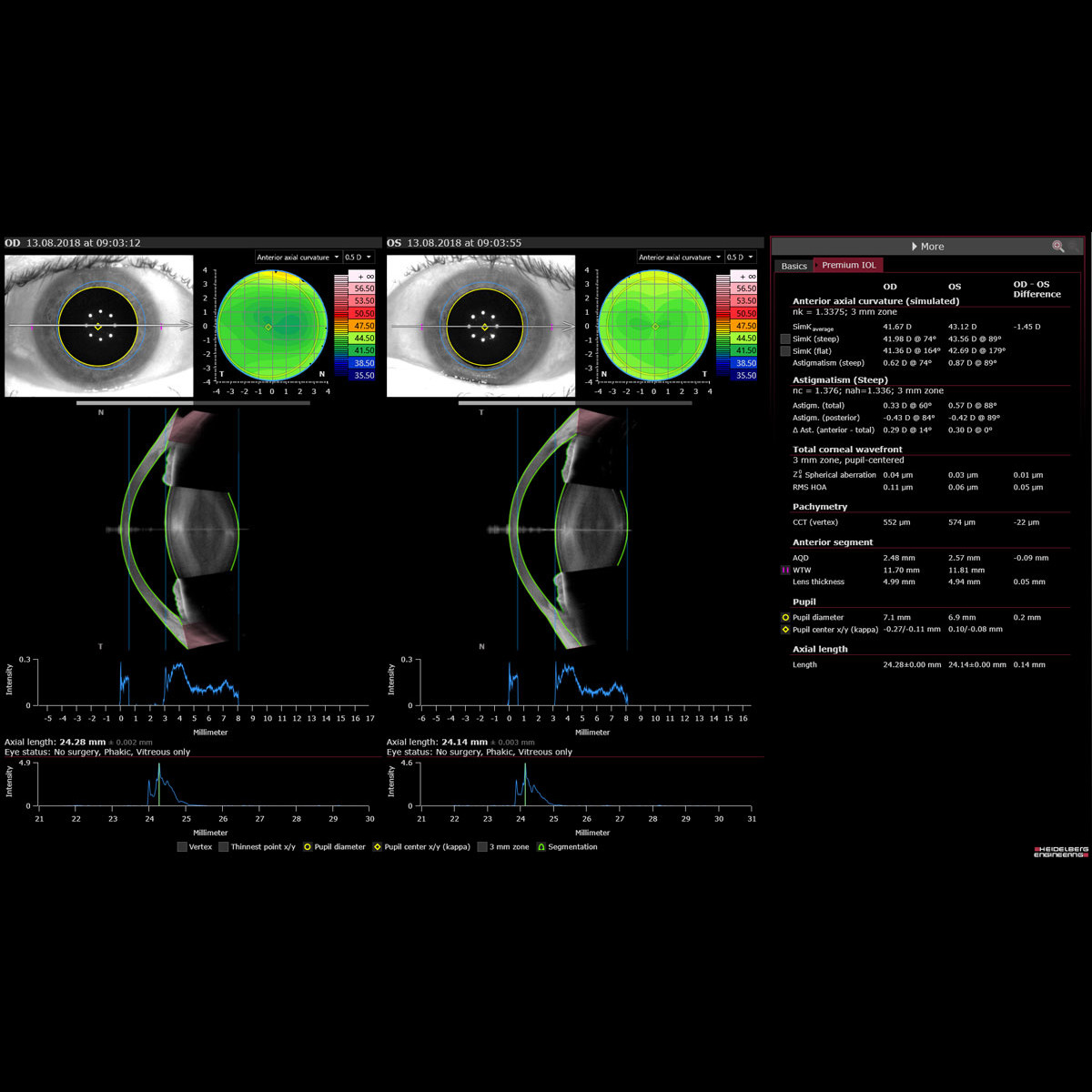
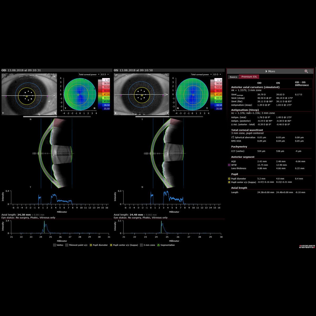
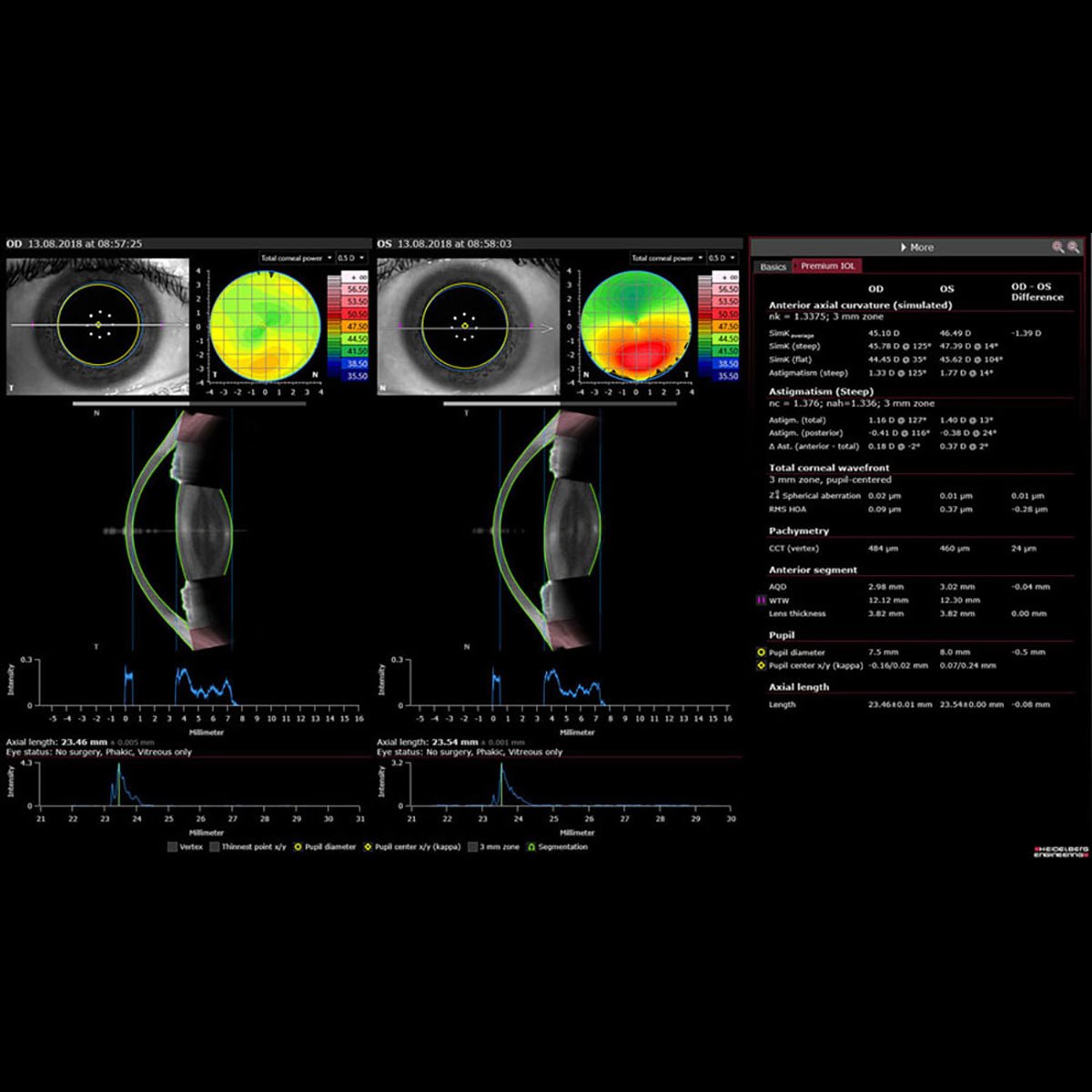
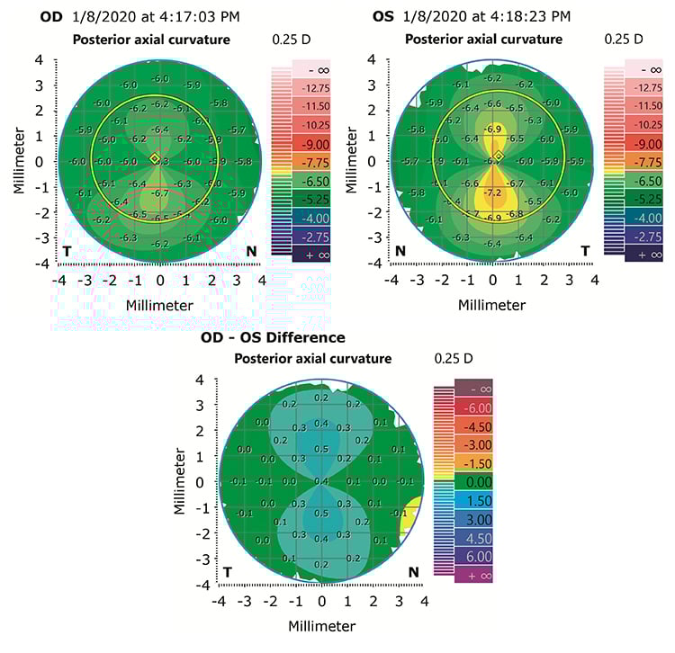
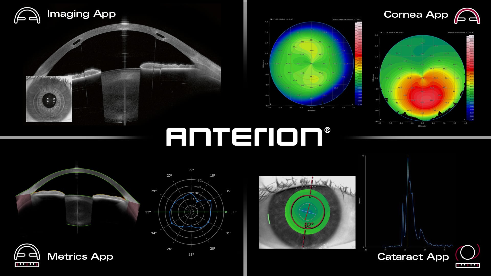
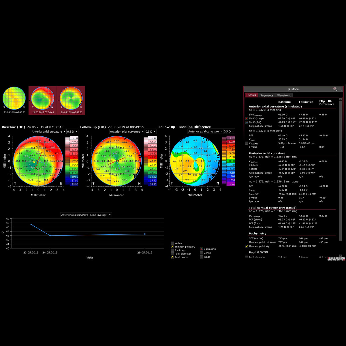
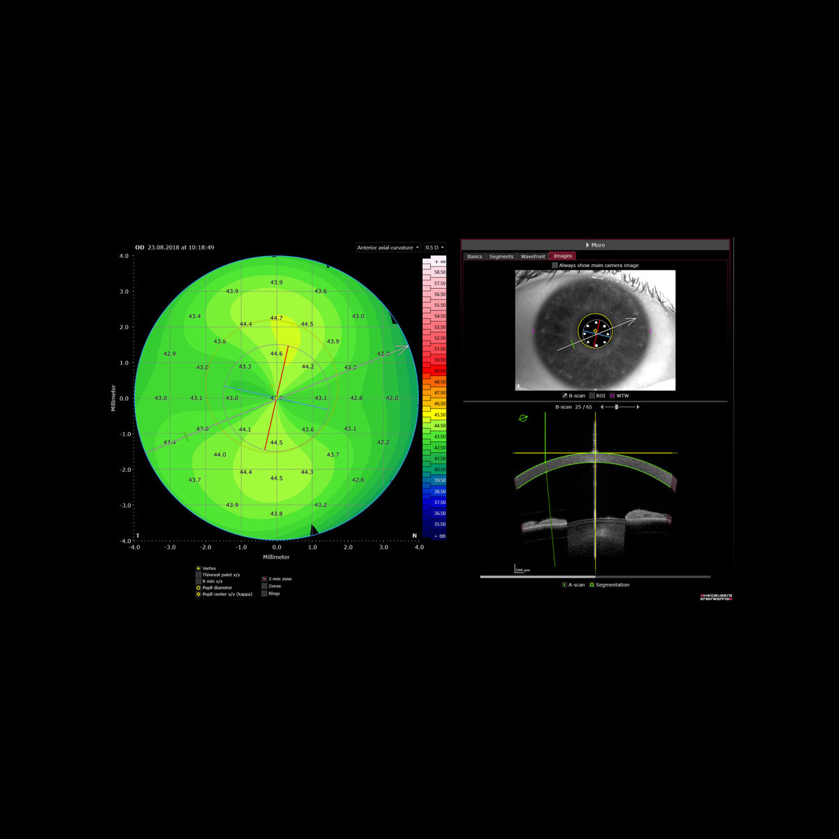
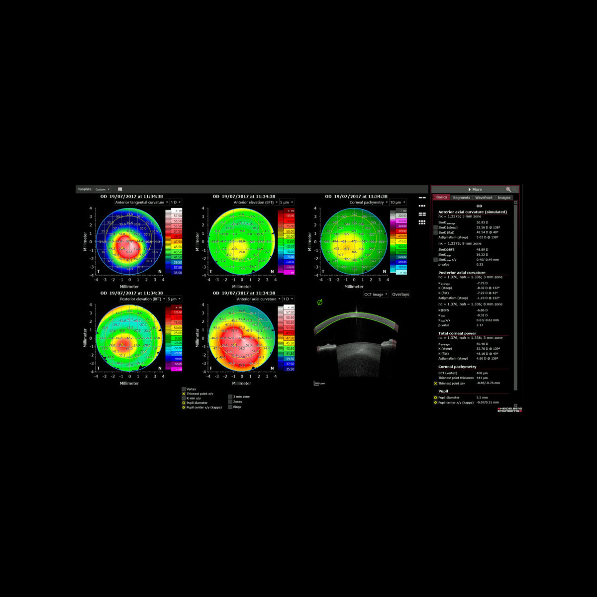
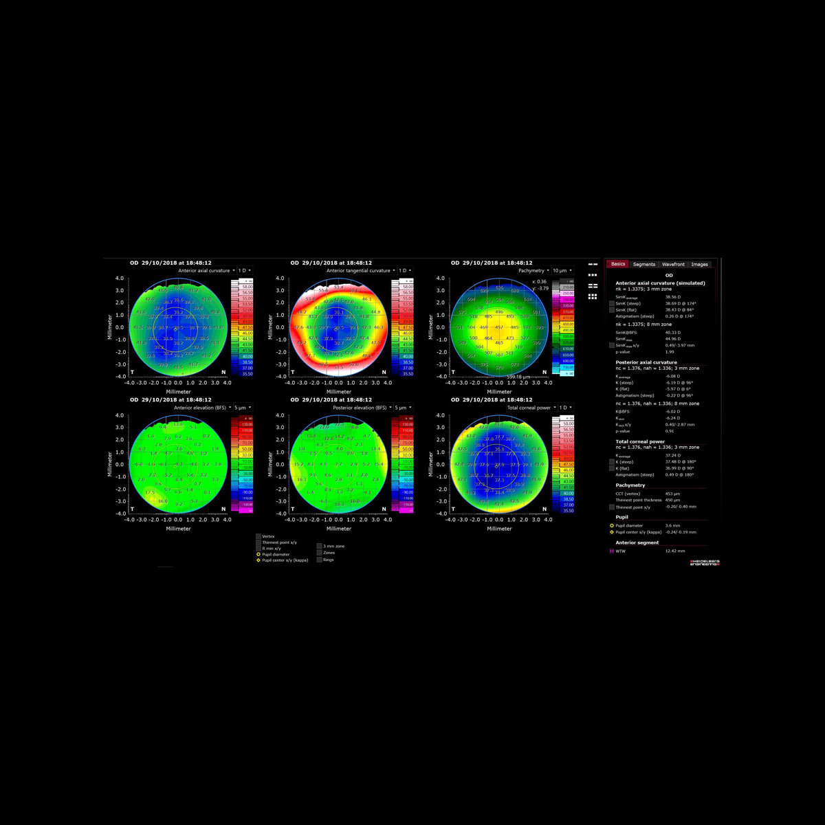
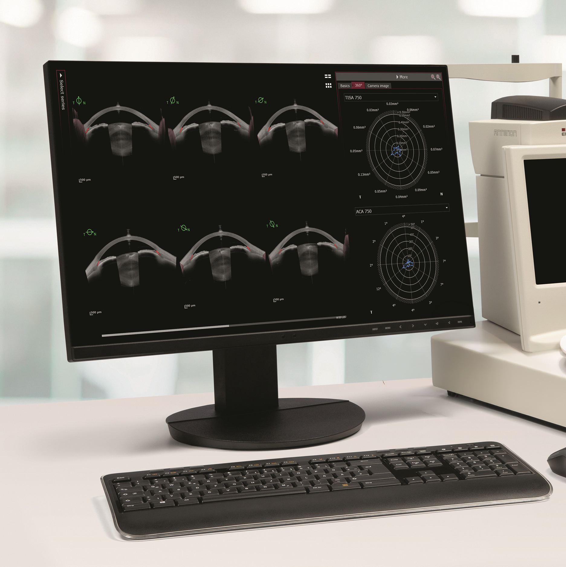
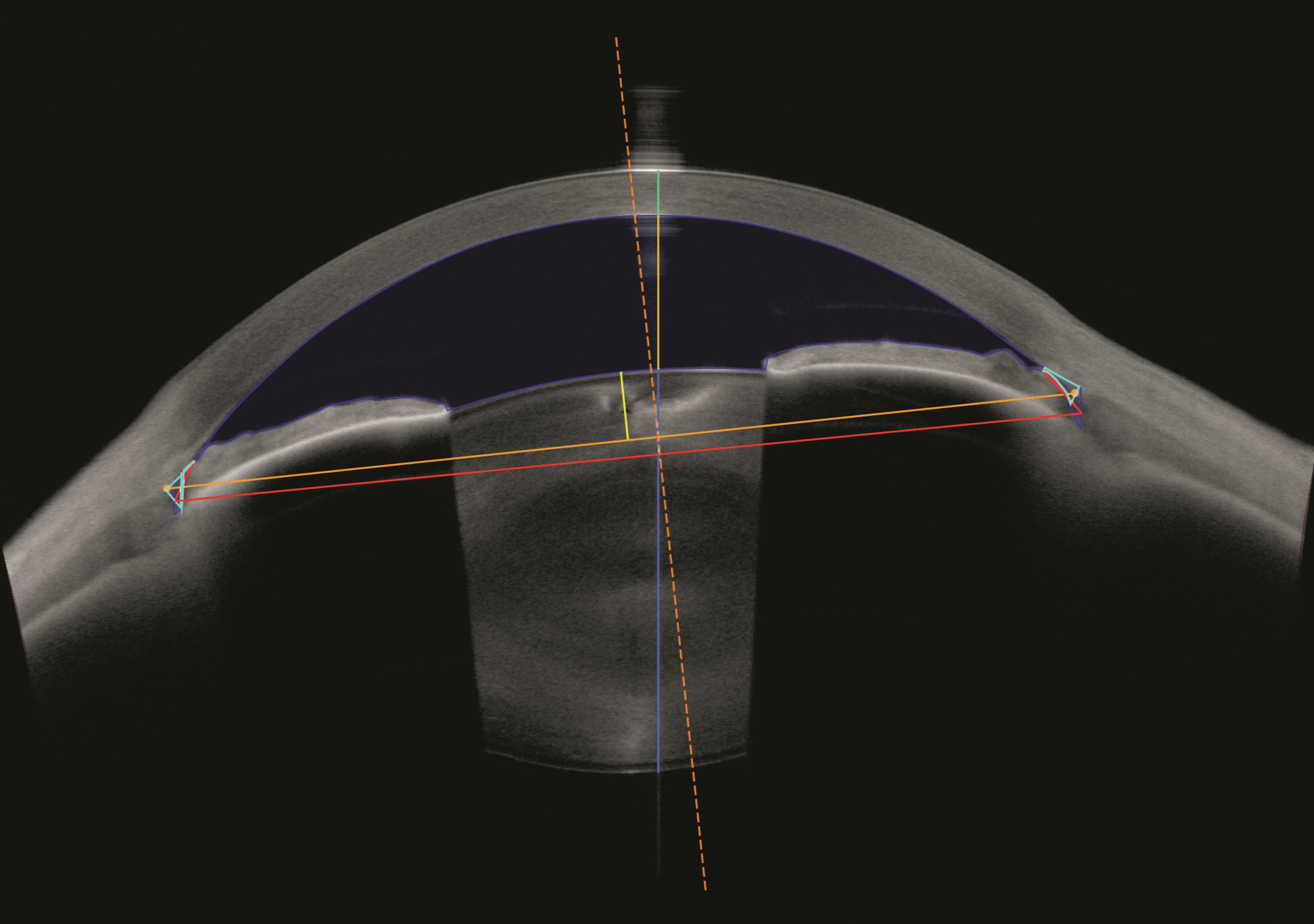
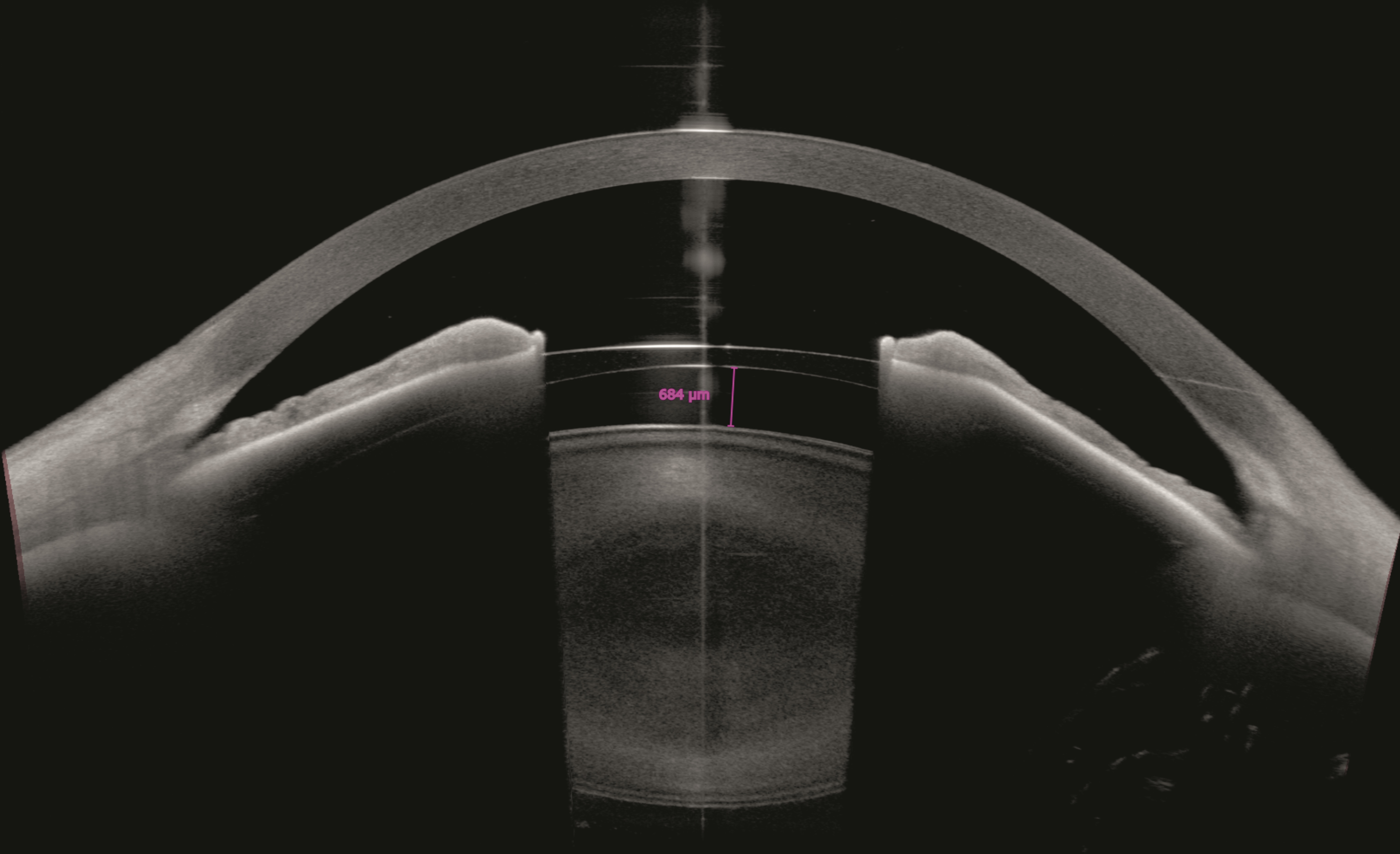
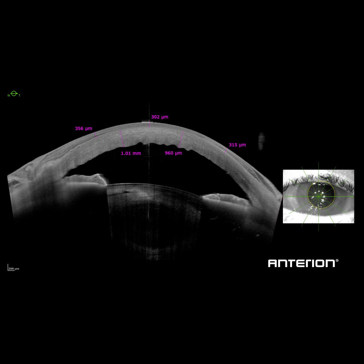
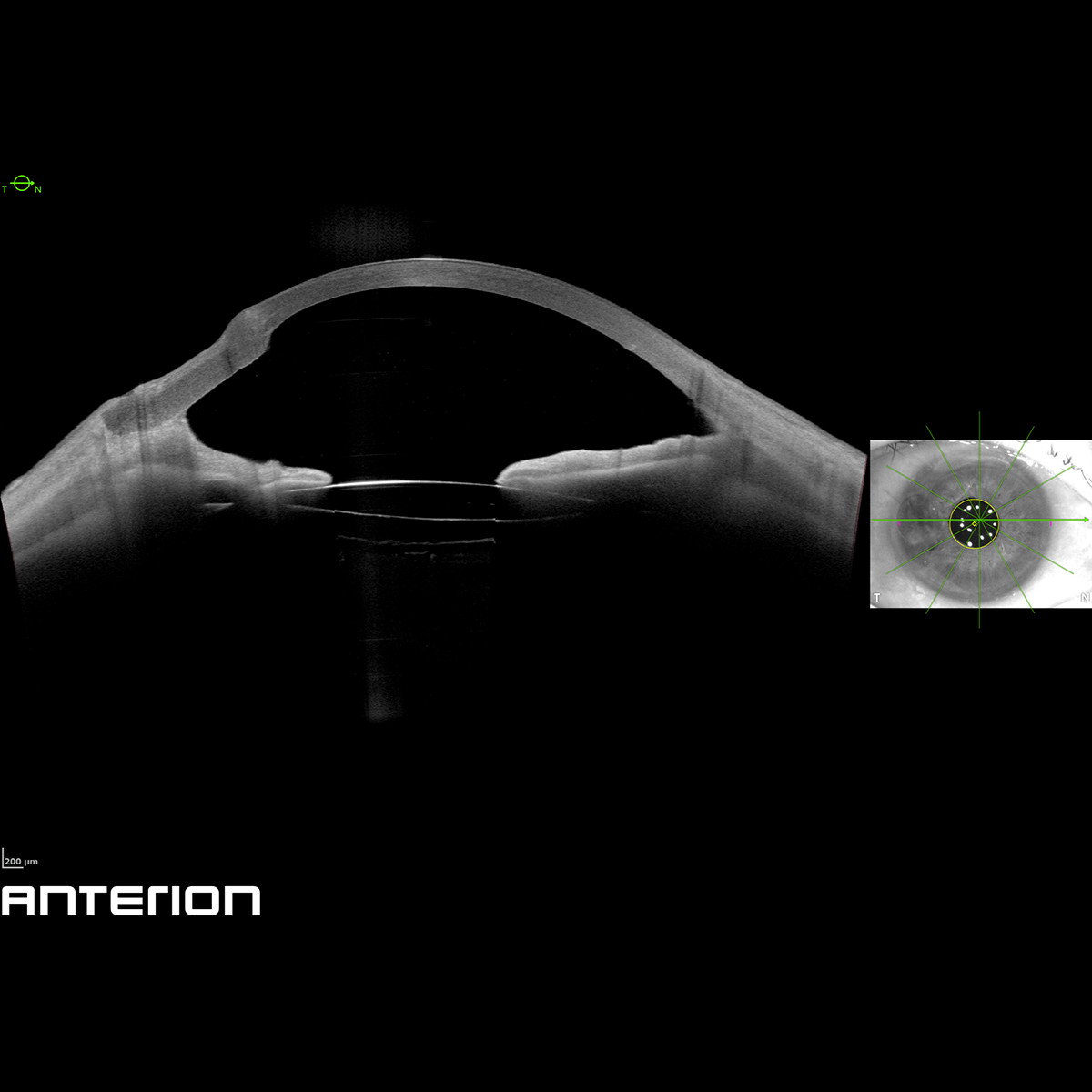
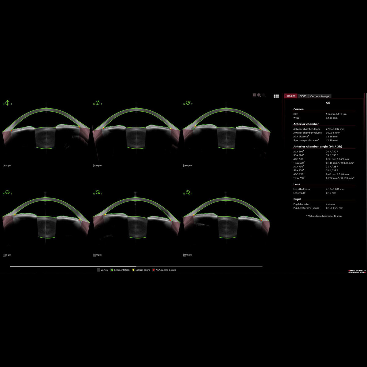
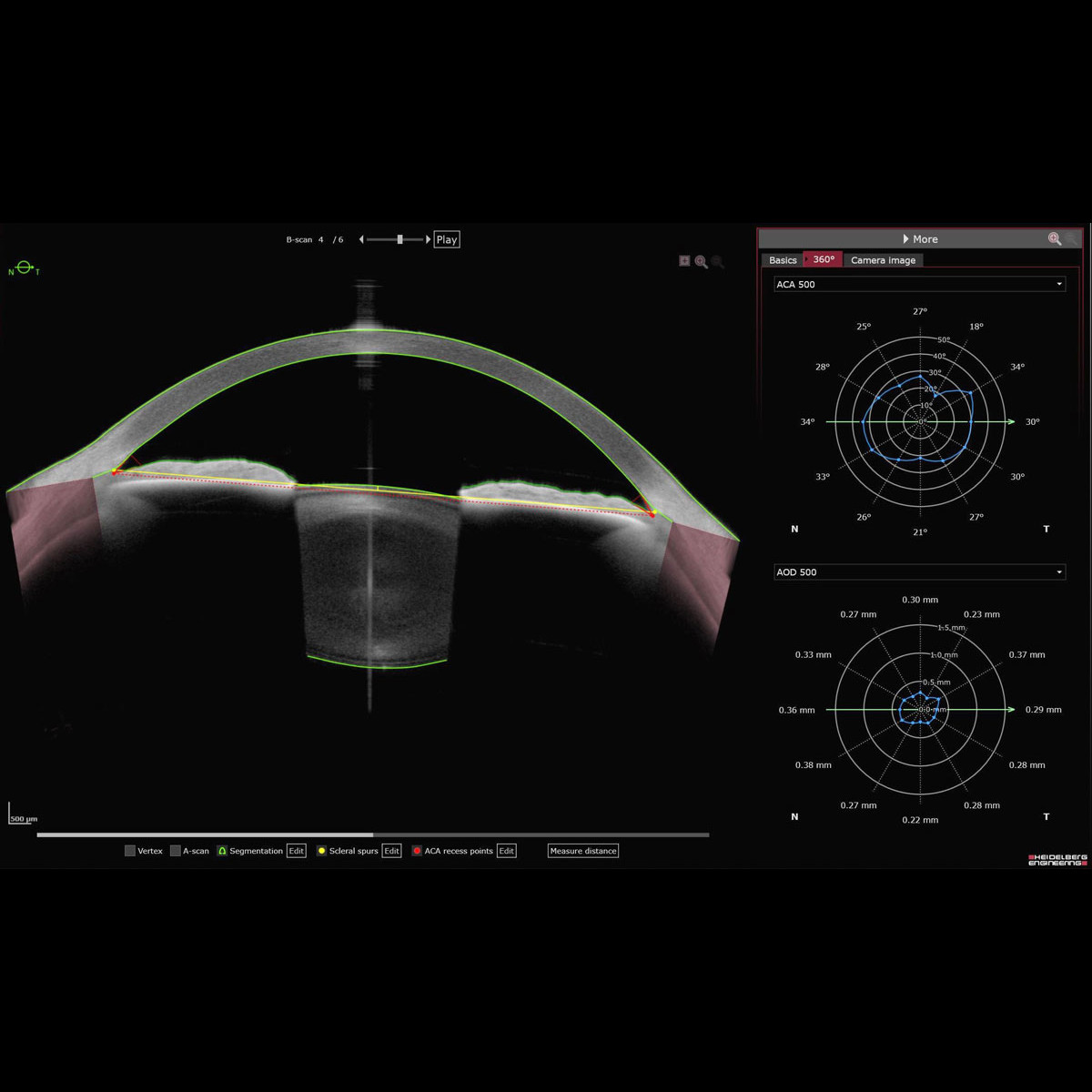
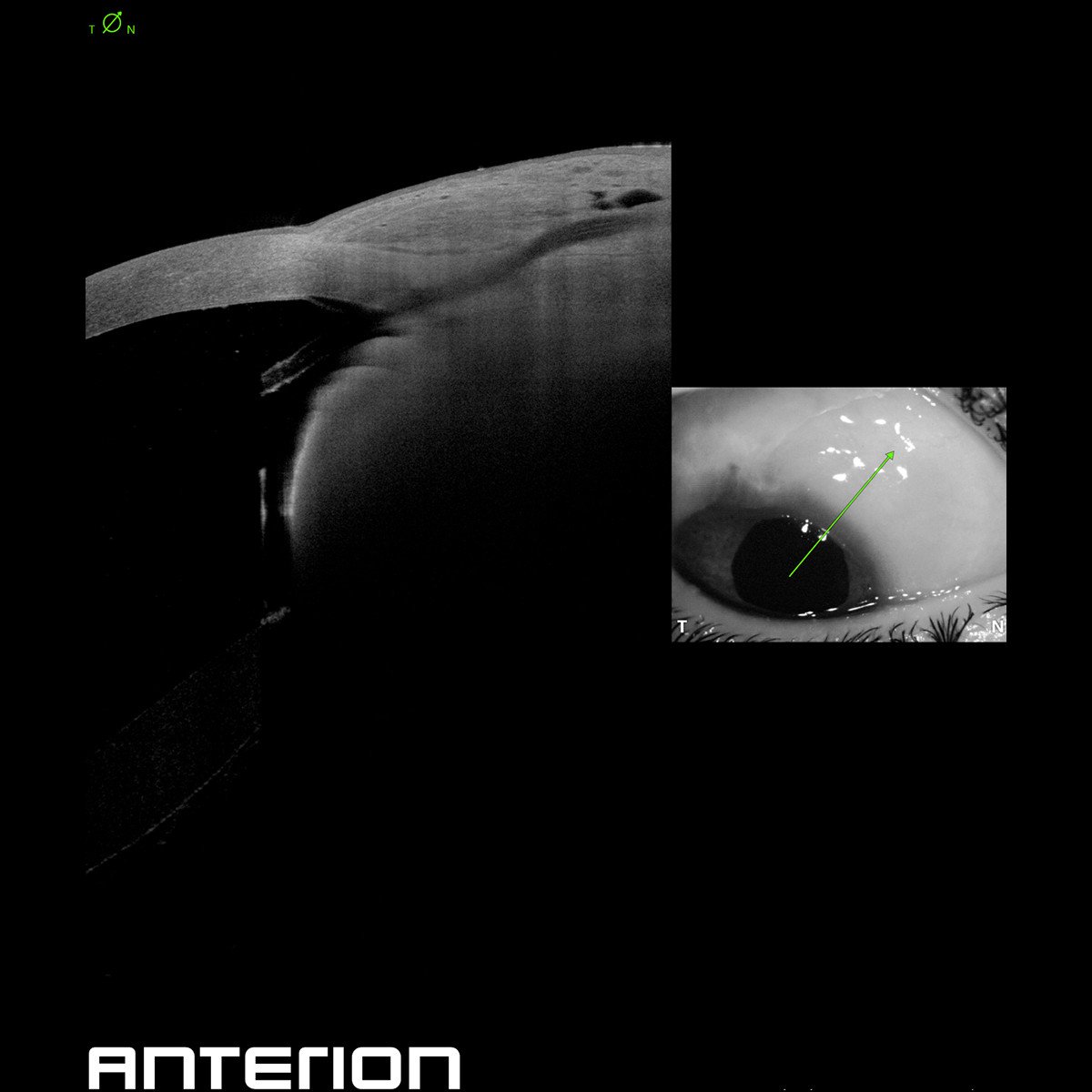
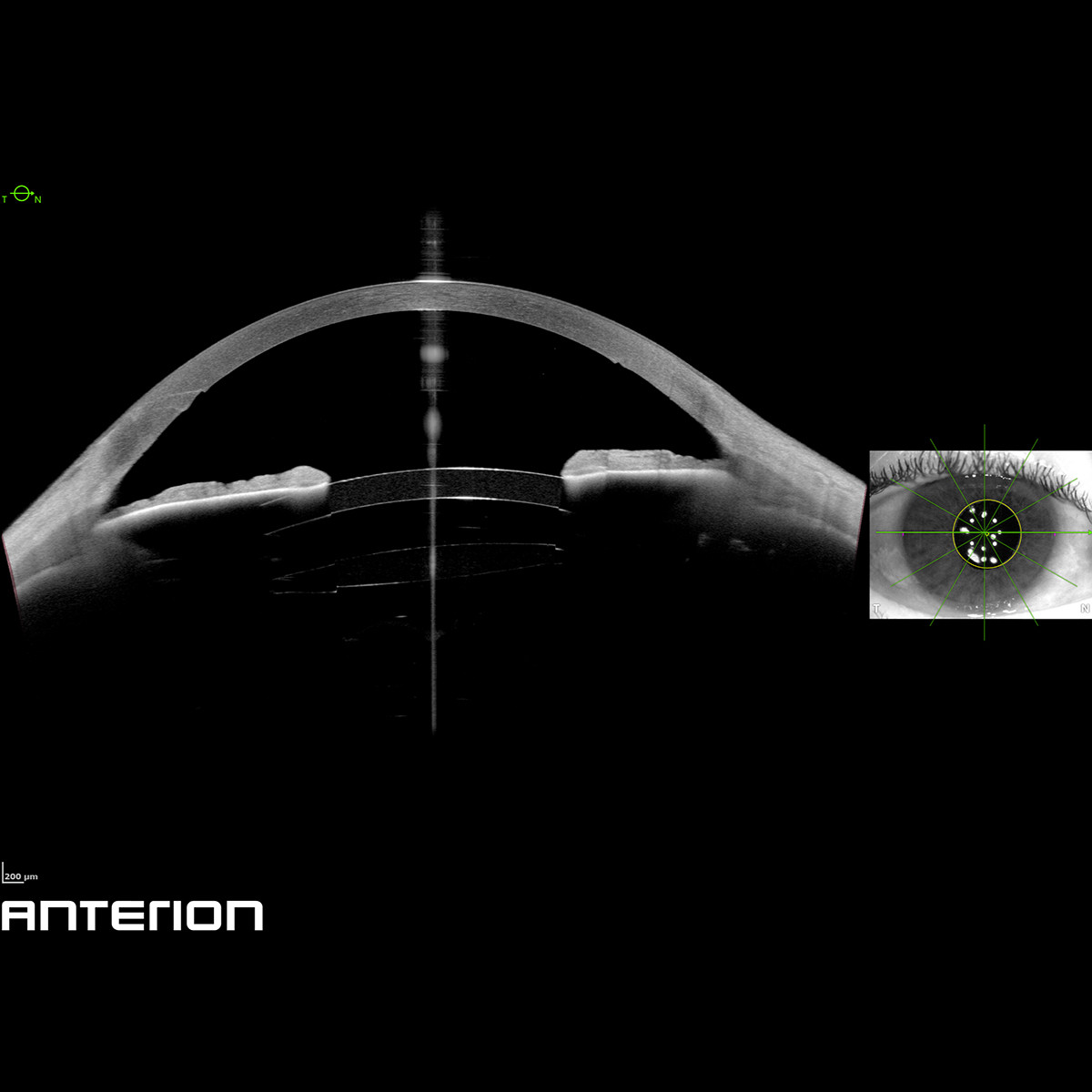
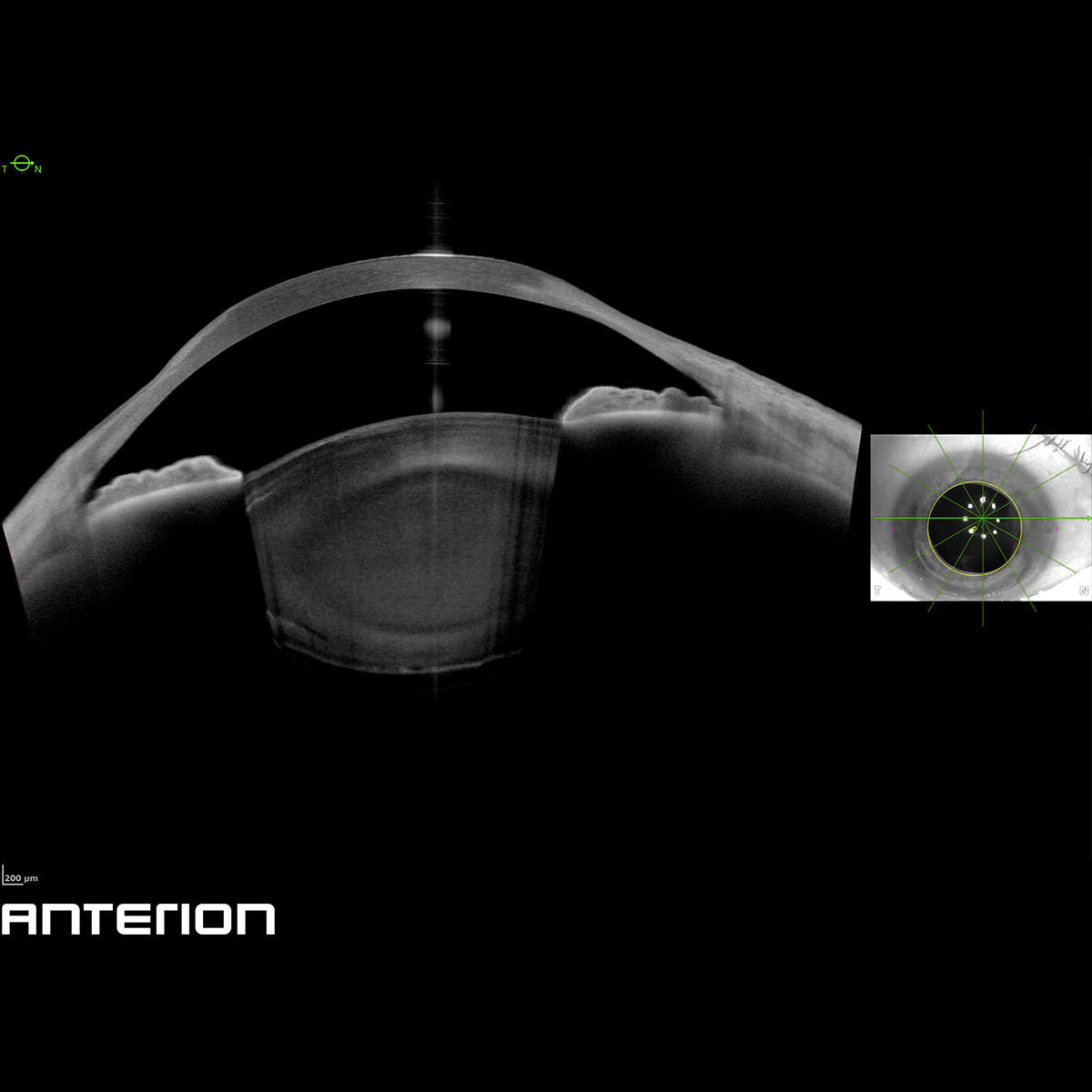
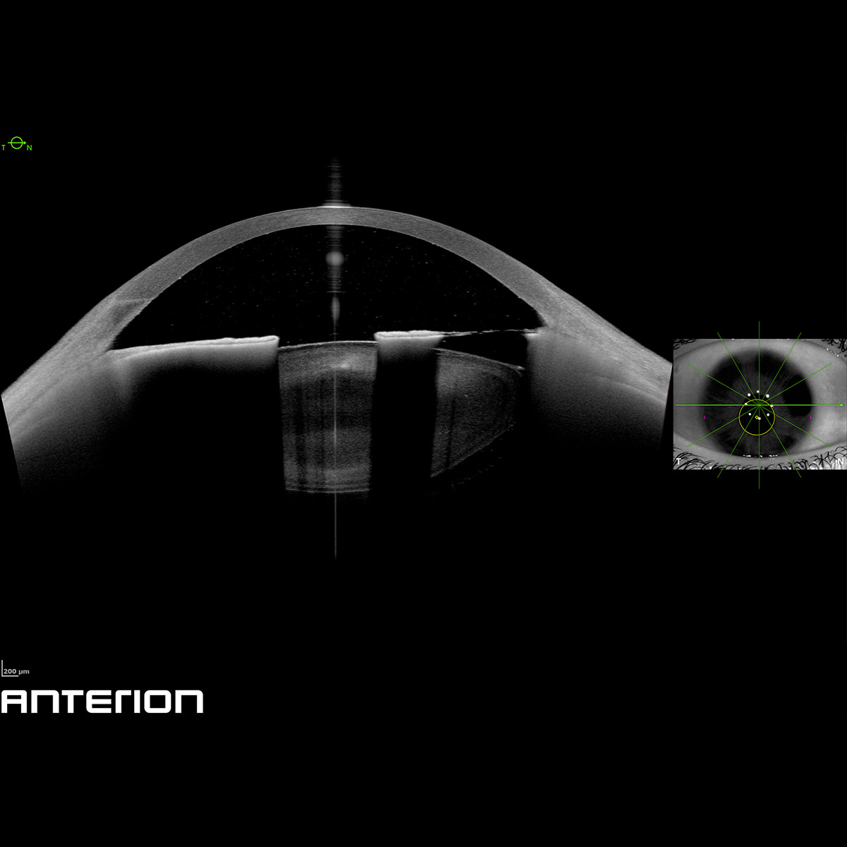
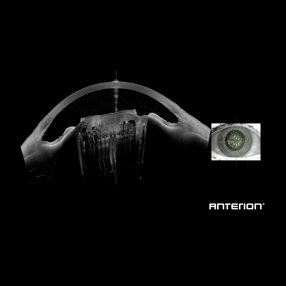
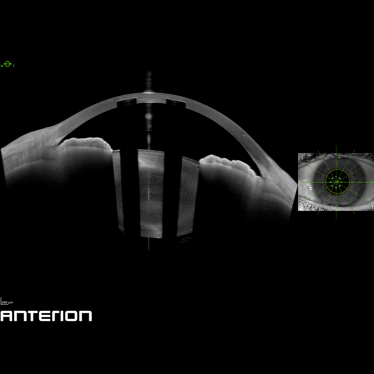
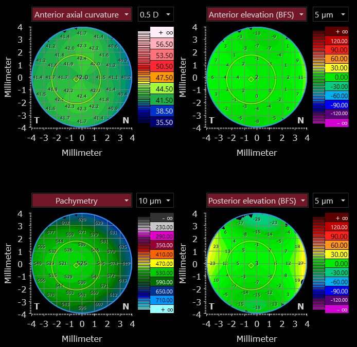
As the definitive toolbox for cataract surgery planning, ANTERION® acquires precise biometric measurements, as well as total corneal power, all with optimised OCT technology. This helps to improve your clinical decision-making, even in the most challenging cases. The OCT images will help you confirm your measurements, resulting in fewer assumptions.

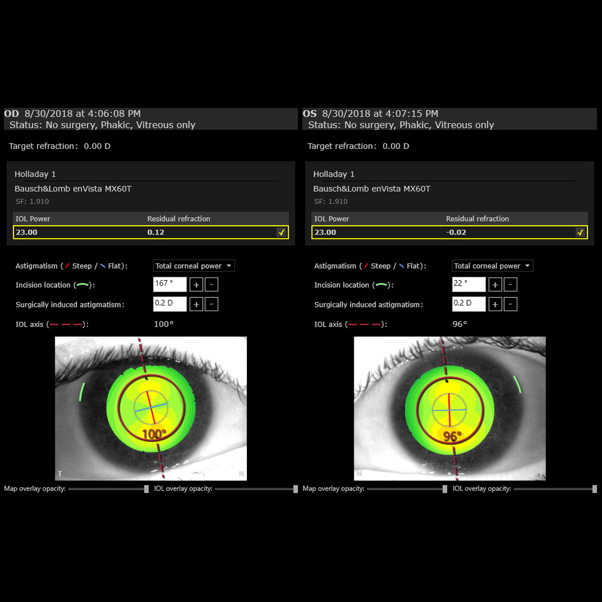
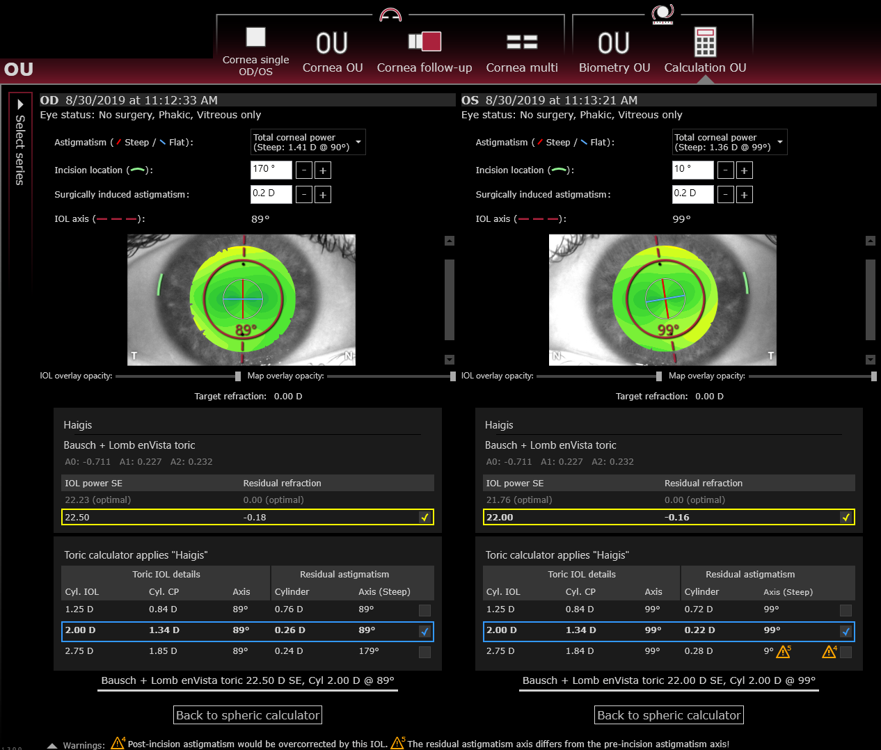
The Cataract App offers spheric and toric IOL calculation as well as raytracing applications and provides the necessary data to calculate the most suitable IOL for your patient, without transferring data or changing devices.
As a connected solution, the data can be exported to DICOM-compatible systems for efficient surgical planning.

Cornea App

Gain a complete view of corneal geometry – with curvature, elevation, pachymetry, total corneal power, epithelial thickness and ectasia assessment. The Cornea App supports confident diagnostics, surgical planning, and follow-up of corneal changes.

1 / 3

Comprehensive Corneal Tomography and Customised Cornea Analysis

The ANTERION® Cornea App provides a full diagnostic suite for analysing corneal geometry using swept-source OCT. It combines curvature and elevation maps of anterior and posterior surfaces with pachymetry and corneal wavefront data. High repeatability and customisable layouts allow clear evaluation of asymmetry, ectasia, disease progression, and surgical outcomes. Additional applications such as epithelial thickness mapping and the SCORE Analyser* for assessing risk of ectasia enhance early detection of corneal irregularities. 

*Powered by Dr. Gatinel and Dr. Saad for AInsight

Metrics App

Visualise and quantify anterior chamber structures in a 360° view. The Metrics App provides automatic and customisable measurements for comprehensive anterior chamber and angle evaluation.

1 / 4

Quantitative Analysis of Anterior Chamber and Angle Parameters

The ANTERION® Metrics App combines clear swept-source OCT imaging with precise quantification of anterior chamber parameters. Automatic scleral spur detection enables immediate calculation of parameters such as anterior chamber volume and angle parameters, lens thickness and vault.

Measure any region of interest directly on the image with refraction-corrected free-hand measurements for customised evaluation. You receive precise, reproducible measurements and anterior chamber angle analysis that can complement gonioscopy and support glaucoma management, cataract surgery, or phakic IOL planning.

Imaging App

Visualise the anterior segment in exceptional detail – from the anterior surface of the cornea to the posterior surface of the lens. The Imaging App provides sharp, revealing OCT images that form the foundation of every ANTERION® examination and workflow.

1 / 3

High-resolution Imaging that Supports Diagnostic Confidence

As the core of the ANTERION® Platform, the ANTERION® Imaging App delivers high-resolution swept-source OCT images of the full anterior segment. Clinicians can visualise surgical outcomes such as keratoplasty, implanted IOLs and corneal rings. The detailed visualisation of cornea, sclera, ciliary body, and rectus muscles provides valuable insights for diagnosis and follow-up of anterior segment conditions.