You are currently on the International (English) version.

Looks like you are in United States.

Switch to local content?

Faster. Reliable. Patient-centered. Uncover What’s NEW on SPECTRALIS® - Heidelberg Engineering’s Flagship Multimodal Imaging Platform

Heidelberg Engineering announces the latest software release for its flagship SPECTRALIS® multimodal imaging platform, introducing enhanced workflows, high-quality imaging, and strengthened diagnostic confidence to support clinicians in improving patient care.

Heidelberg Engineering announces the latest software release for its flagship SPECTRALIS® multimodal imaging platform, introducing enhanced workflows, high-quality imaging, and strengthened diagnostic confidence to support clinicians in improving patient care.

Since its introduction, Heidelberg Engineering’s SPECTRALIS® platform has been a benchmark in multimodal retinal imaging – trusted in eye clinics, hospitals, and research centers around the world. With the new release on SPECTRALIS®, the company takes the next step in imaging evolution: enhancing workflow efficiency, imaging speed, and diagnostic confidence. This next expansion of the SPECTRALIS® multimodal functionality adds a suite of new tools, from Green Autofluorescence to faster OCT Angiography Module and flexible reporting options, that build on the platform’s well-established DNA in enhancing diagnostic confidence and continuously improving patient care. The newest features are only compatible with HEYEX 2.

Anticipated by SPECTRALIS® users worldwide, the new release reinforces the platform’s leading role in posterior segment imaging. It delivers valuable new features for generalists and specialists in both retina and glaucoma, designed to improve clinical workflows and enable more precise assessments.

Faster and Personalized Imaging

RETINA: The next generation of features within the retina suite help expand clinical insight by empowering clinicians to tailor imaging protocols to patient needs. 

With the introduction of a new and faster scan speed, SPECTRALIS® with SHIFT technology expands its scan speed portfolio. The addition of 250 kHz to the existing 85 kHz and 125 kHz scan speeds allow faster OCT angiography image acquisitions and an improved OCTA workflow. Supported by enhanced TruTrack Active Eye Tracking, 250 kHz helps ensure reliable image quality throughout the examination. 20 kHz scan speed in the SHIFT portfolio remains the ideal scan speed for imaging challenging patients, when a higher signal-to-noise ratio is required, for e.g., in eyes with media opacities.

A clinical comparison of 250 kHz scan speed with 125 kHz scan speed in OCTA imaging, with both speeds powered by the enhanced TruTrack Active Eye Tracking technology, suggests 43%* reduction in acquisition time with the faster speed. Shorter examination times greatly benefit patients who struggle to maintain fixation, particularly elderly, pediatric, or low-vision individuals.


 

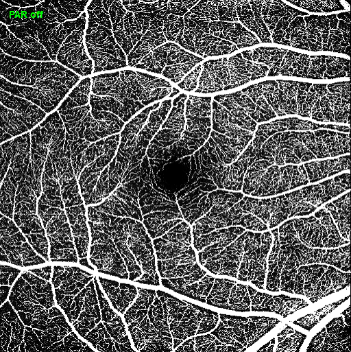
Transverse OCT angiography image (superficial vascular plexus) of a healthy eye using 250 kHz scan speed with enhanced TruTrack Active Eye Tracking

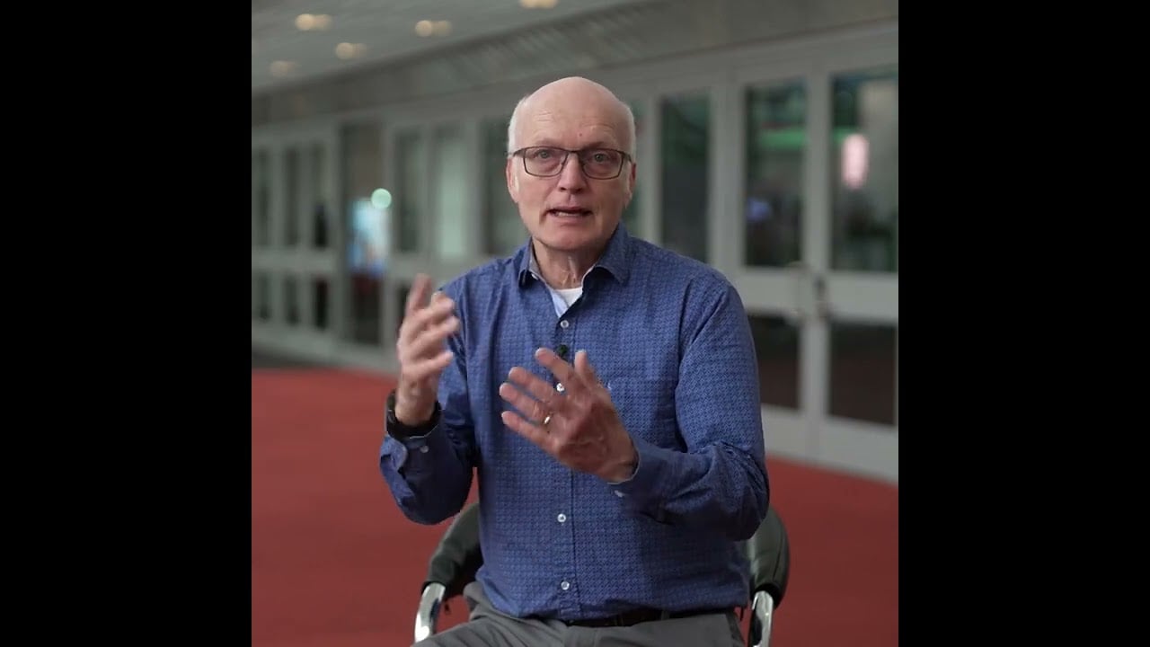
“The benefit of the (enhanced) TruTrack Active Eye Tracking and high-speed OCTA is that the patient spends less time getting imaged and we can image more patients per SPECTRALIS®. The patients come every 4-6 weeks for their treatment and it is always a burden for them to come for longer OCTA examinations.”

Dr. Dirk-Uwe Bartsch, California, USA
Confident Diagnostic Decisions

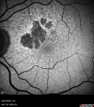
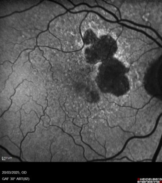
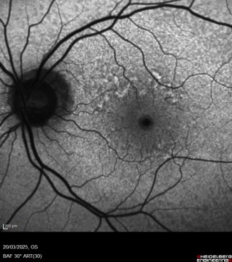
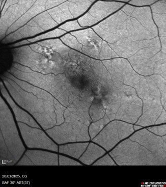
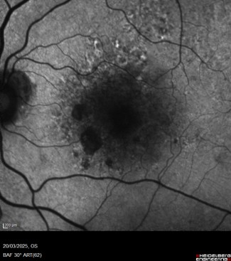
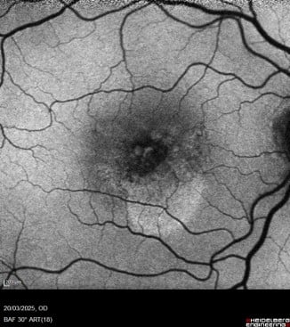
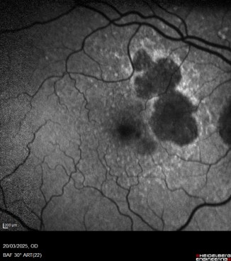
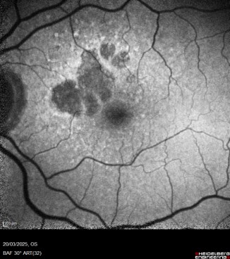
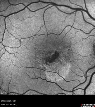
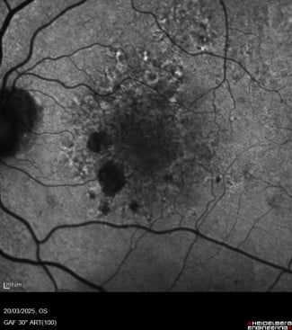
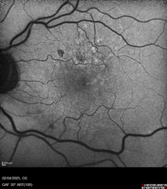
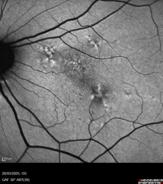
RETINA: The expanded scope of SPECTRALIS® also includes advanced imaging capabilities that allow deeper insights into retinal and optic nerve health. The Green Autofluorescence Module, using 518 nm excitation light, improves visualization of foveal sparing in macular atrophy and supports the assessment of lipofuscin distribution and retinal pigment epithelium loss, which is valuable in managing conditions such as geographic atrophy (GA) and Stargardt’s disease. Combined insights with the BluePeak Module strengthen the SPECTRALIS® portfolio of Multimodal Imaging.


“The Green Autofluorescence really depicts how you see the fovea and how the atrophy can be seen inside the fovea, which is very important to quantify the patient’s geographic atrophy lesions…it is recommended to do both at the same time (BAF+GAF). From the patient’s perspective, it is very important that the examinations are fast and precise so to get the most out of your time with the doctor and the green autofluorescence gives you a good overview of how the foveal involvement is described.”

Gregor Reiter, Vienna, Austria
1 / 13

Further, the Low Luminance Autofluorescence feature allows clinicians to adjust the intensity of blue laser, enabling high-quality imaging at lower illumination levels when using autofluorescence or reflectance imaging modalities. 

The addition of Dense ART scan patterns (ART values of up to 100) for OCT angiography imaging allows averaging over a small area for the visualization of specific regions of interest and more detailed interrogation of both structure and flow in vascular pathologies. 

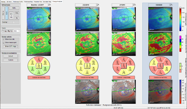
GLAUCOMA: The updated Change Analysis function for the Posterior Pole scan in the SPECTRALIS® Glaucoma Module Premium Edition offers increased flexibility in analyzing the Ganglion Cell Complex (GCC). Clinicians can now segment or combine layers of the GCC to visualize disease progression over time, conveniently displayed in a single, comprehensive view. 

The Change Analysis tab for the Posterior Pole scan with SPECTRALIS® Glaucoma Module Premium Edition

RETINA and GLAUCOMA: Users can create additional custom scan presets directly in the acquisition module. Each preset stores defined parameters, allowing teams to standardize imaging protocols across clinics, research sites, or patient groups.

The multiselect print function reduces manual selections in the report printing process that cost time and effort by creating and combining all desired reports into one. It’s possible to set templates as default for other patients and fully document patient visits. Users can now experience a simplified documentation process, reduced administrative time, and consistent reporting practices in clinics due to these configurations. 

The newest SPECTRALIS® features are designed to help users get even more from multimodal imaging. We’re excited to see how these innovations continue to support clinical workflow and thereby impact patient outcomes worldwide.
The software is now commercially available. 

Uncover more >>> 

DISCLAIMER: This content may contain product information differing from the current approval status in your country. Please refer to your country-specific website for relevant information about your market.

*Morsy MS, Mehta NN, Ali AL, et al. COMPARISON OF ULTRAHIGH-SPEED 250 kHz OCTA WITH HIGH-SPEED 125 kHz MODE USING HEIDELBERG SPECTRALIS SHIFT: A Paired-Eye Analysis. Retina. 2026;46(5):829-835. doi:10.1097/IAE.0000000000004751