













Analyze the Cornea at the Cellular Level
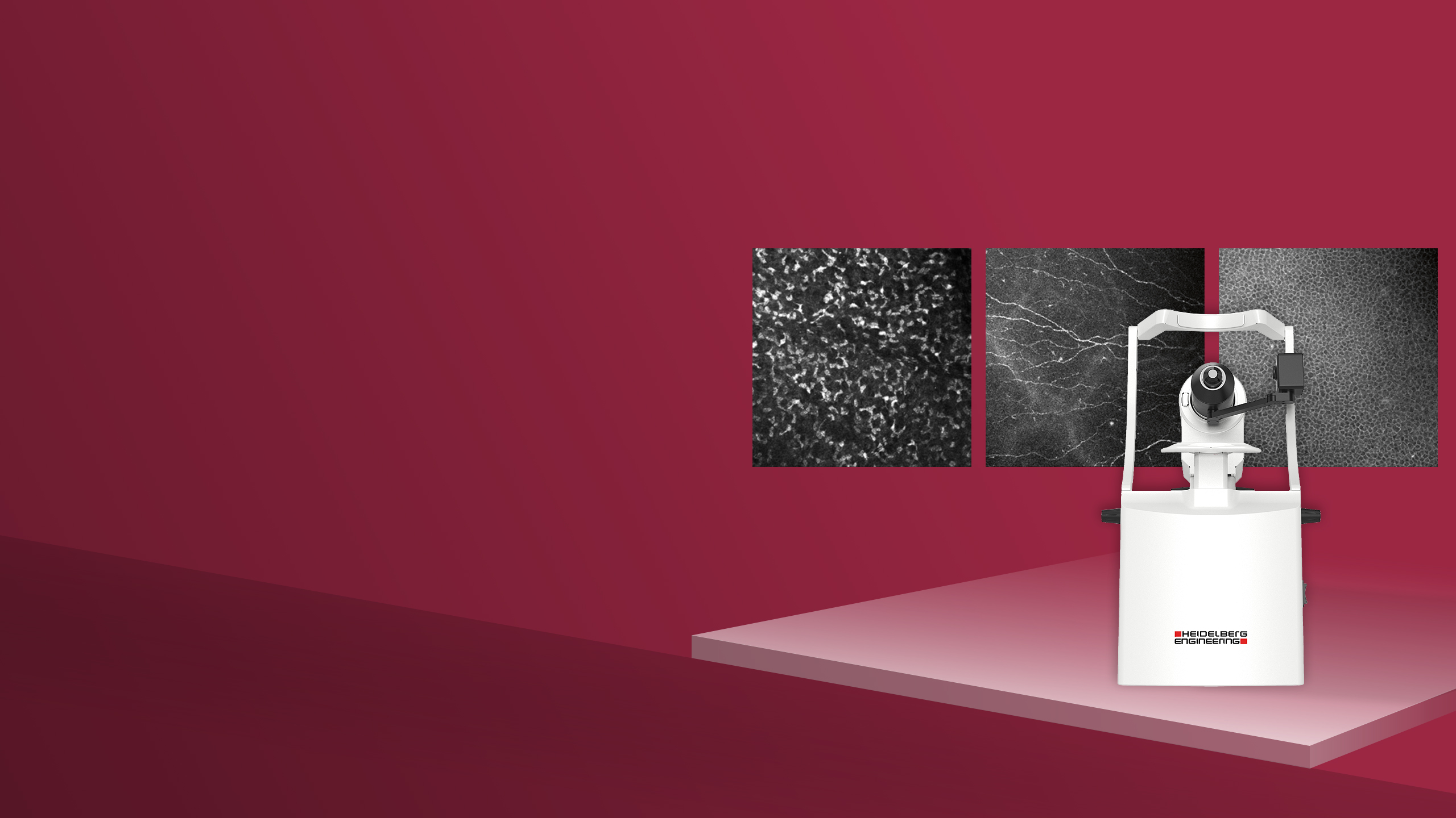
Heidelberg RCM allows you to acquire unique en face images of corneal cells and microstructures non-invasively. Examine keratocyte subpopulations, visualize the fine details of the subbasal nerve plexus and assess endothelial cell morphology.
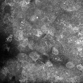
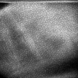
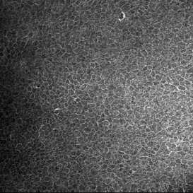
Epithelium
Visualize epithelial cells and assess their morphology at a selected imaging depth.
1 / 3
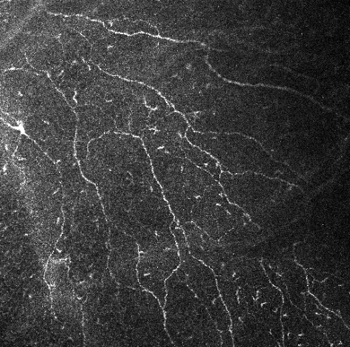
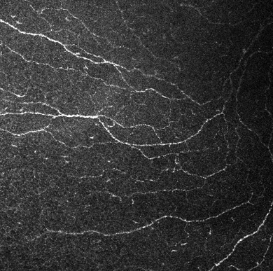
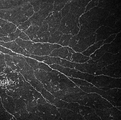
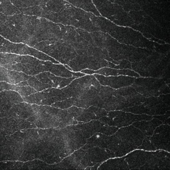
Subbasaler Nervenplexus
Untersuchen Sie das Netzwerk der Hornhautnerven und prüfen Sie die Nervenmorphologie im Detail.
1 / 4
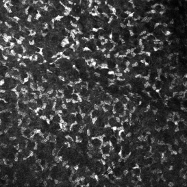
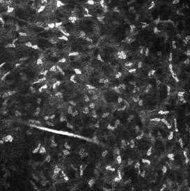
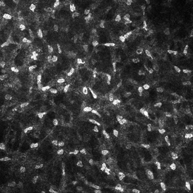
Stroma
Explore stromal microstructure at different imaging depths and visualize the morphology of structures including keratocytes.
1 / 3
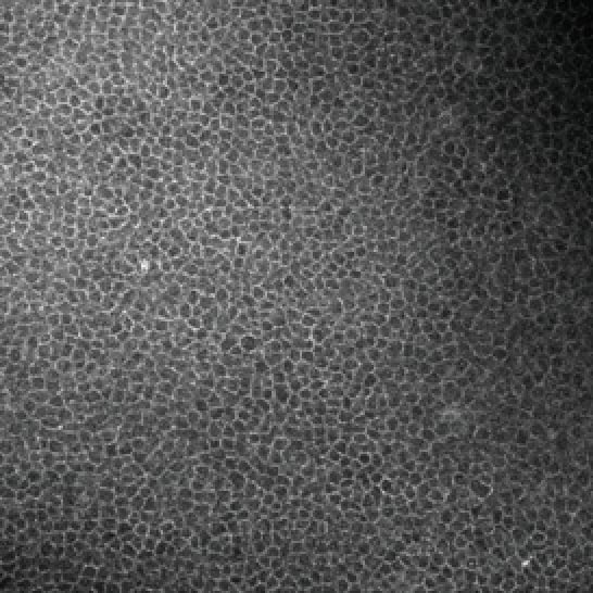
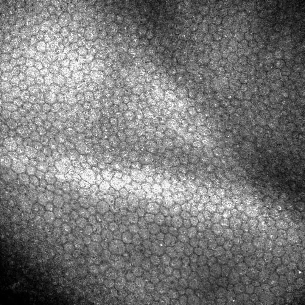
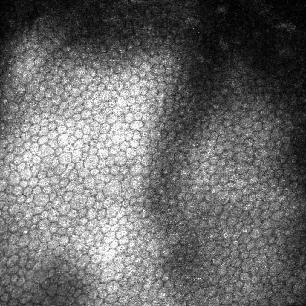
Endothelium
Visualize the corneal endothelium and assess cellular morphology.
1 / 3
Image courtesy: Gaush Meditech