










In Vivo Cellular Imaging Optimized for the Cornea

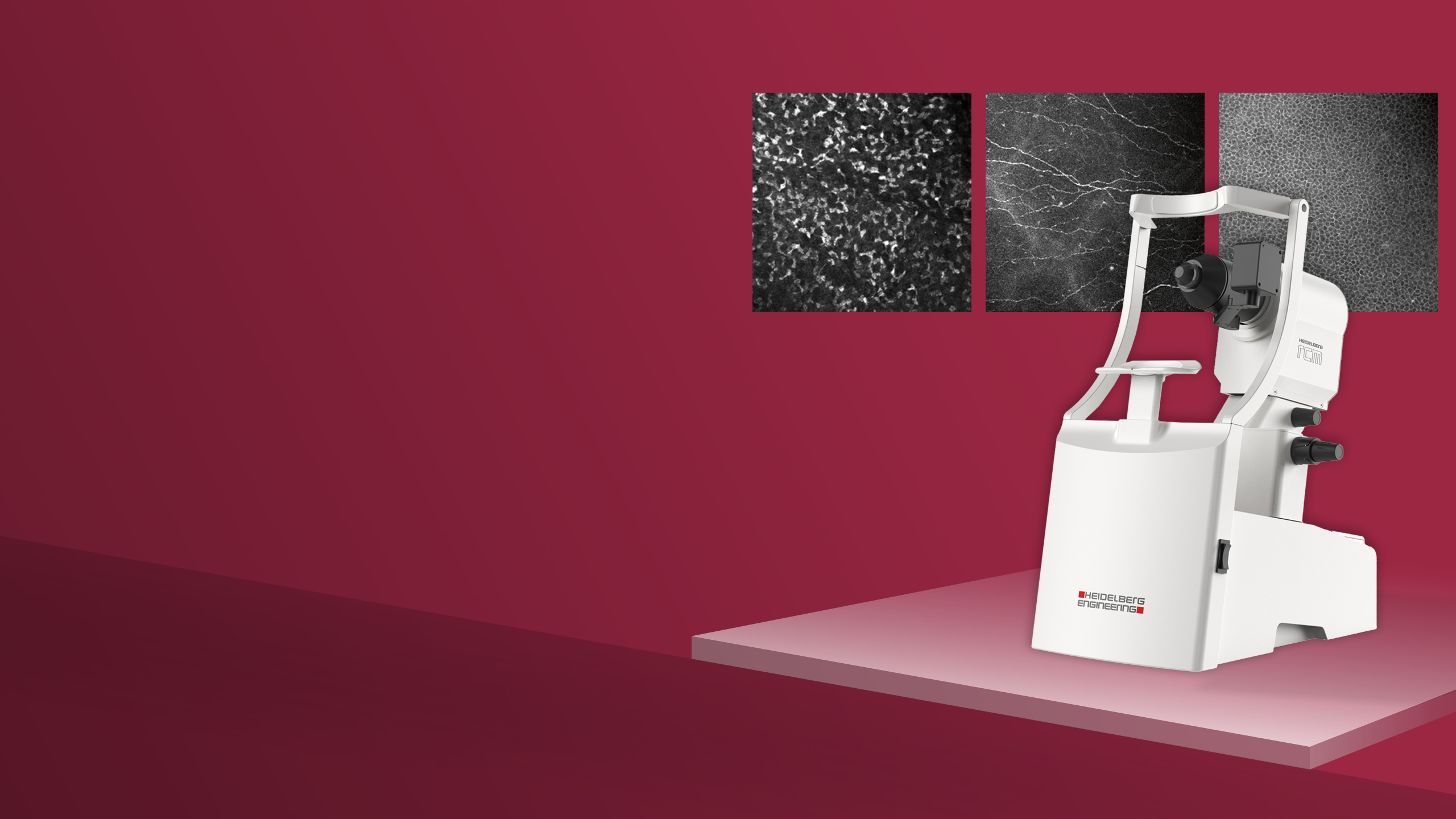
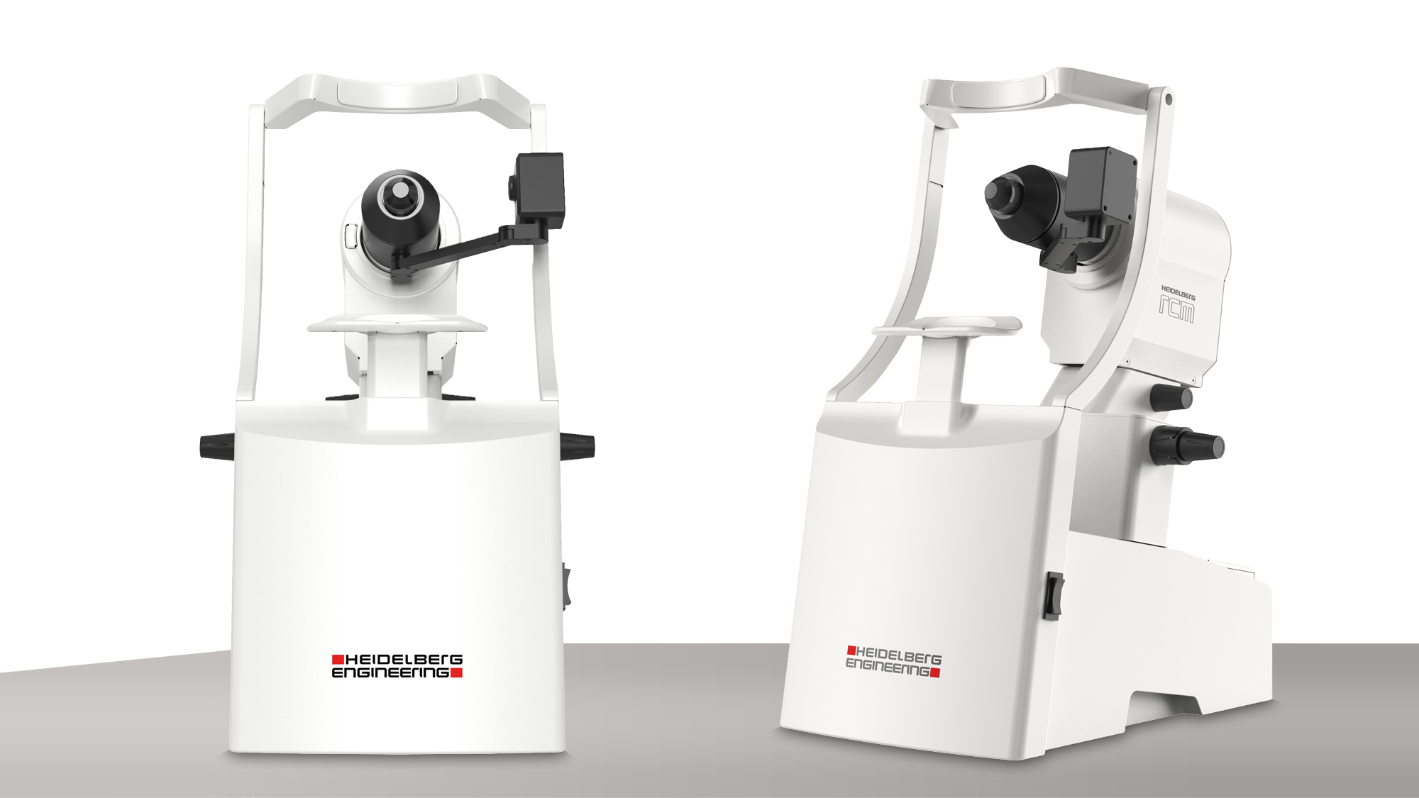
Heidelberg RCM is a compact ophthalmic device that uses confocal scanning laser microscopy for non-invasive imaging of the cornea, providing high-resolution, wide-field microscopic images to examine cellular morphology.

Examine in Vivo. Explore by Layer. Analyze in Detail.

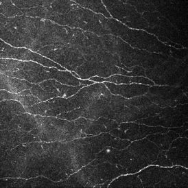
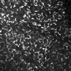
Heidelberg RCM provides detailed en face images of corneal cells and structures without the need for tissue excision.

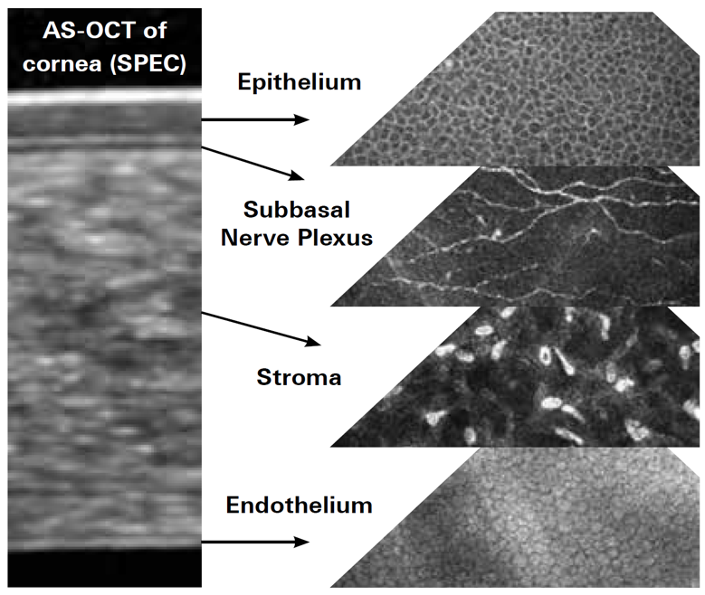
From the epithelium and subbasal nerve plexus to the stroma and endothelium, Heidelberg RCM enables you to explore the cornea at a cellular level and examine morphological changes over time.

Benefits

Corneal Detail to Support Your Assessment

High-resolution en face images reveal corneal cells and structures that cannot be examined in the same detail with conventional clinical imaging. Heidelberg RCM allows you to explore the cornea and supports detailed analysis and follow-up across clinical practice and research. 
1 / 2
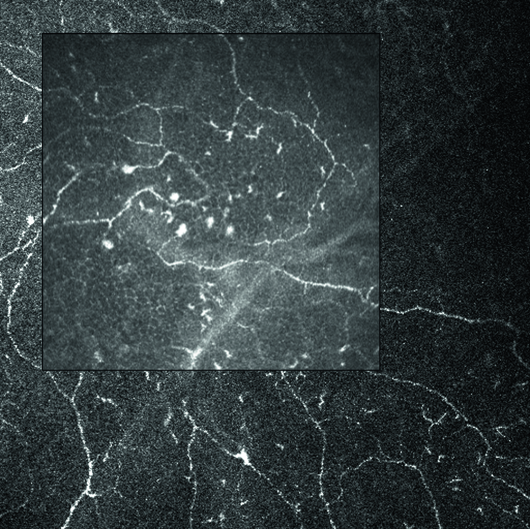
400 x 400 µm field of view vs 600 x 600 µm field of view

See More with a Wider Field of View

Use the 400 × 400 µm field of view for detailed examination or select the optional 600 × 600 µm field of view to visualize a broader area of the cornea within a single image. This flexibility allows you to adapt image acquisition to the structure or area being examined.

Explore the Cornea at Your Selected Depth

Adjust the focal depth to examine the epithelium, subbasal nerve plexus, stroma and endothelium in real-time. This depth-guided imaging enables targeted visualization of the structures and layers relevant to your examination. 

1 / 3

Visual Guidance During Examination

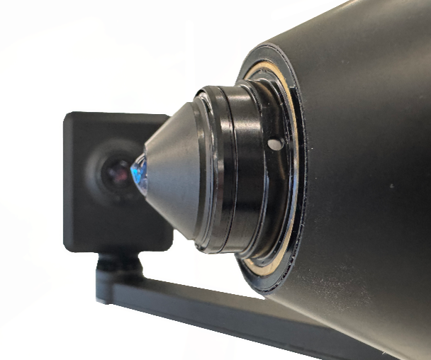
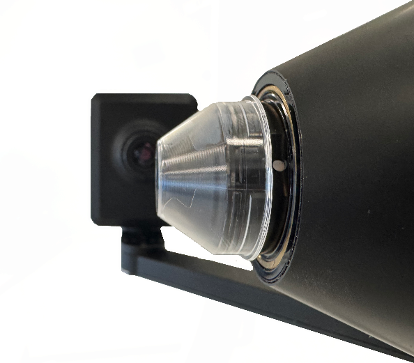
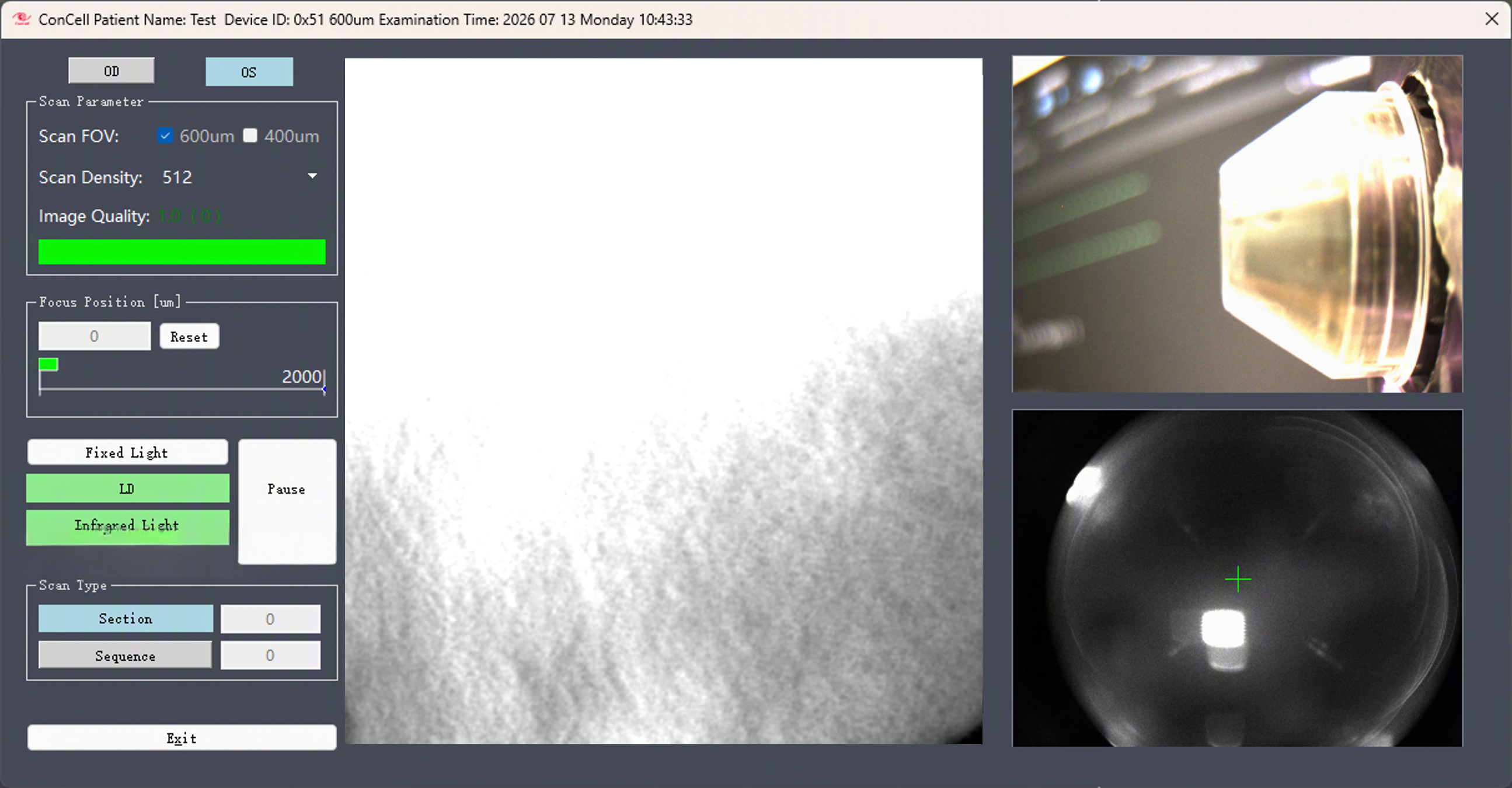
A sterile, single-use TomoCap fits over the microscope lens and provides the interface with the patient’s ocular surface, enabling stable, rotation-free contact during imaging. Two live eye monitoring cameras help you align the cap on the eye and observe its position throughout the examination.

Let’s Talk
about your corneal
imaging needs 

Uncover how Heidelberg RCM can support your clinical practice or research. Contact our team to learn more, discuss pricing, or request a demonstration.  

Subscribe to our newsletter for product news, clinical and research insights, educational resources and upcoming events.