Age-related Macular Degeneration (AMD)

AMD describes age-related changes within the macula—the area of the retina that is important for sharp vision. AMD is the leading cause of central vision loss in people over 50 in Western countries.

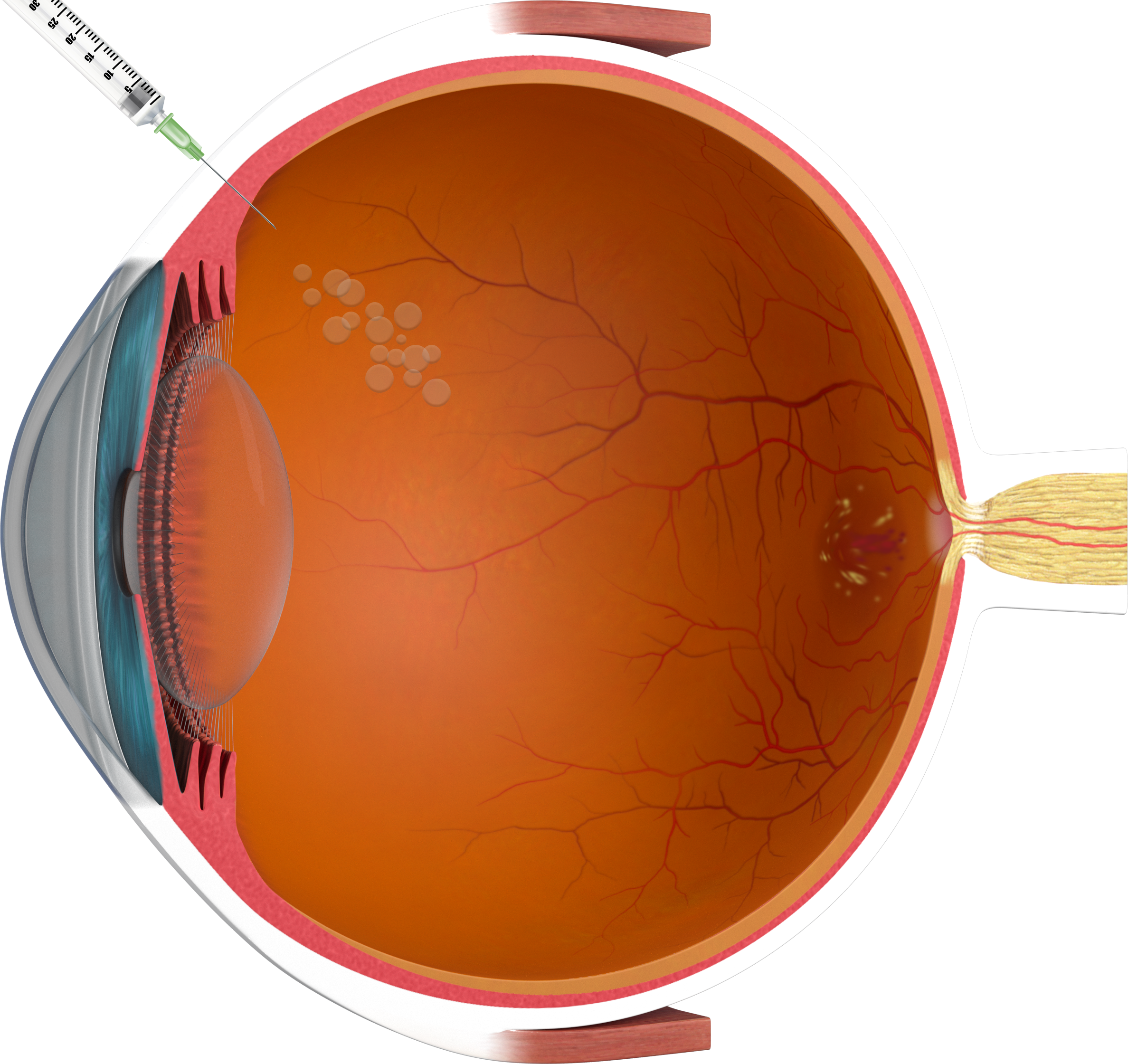
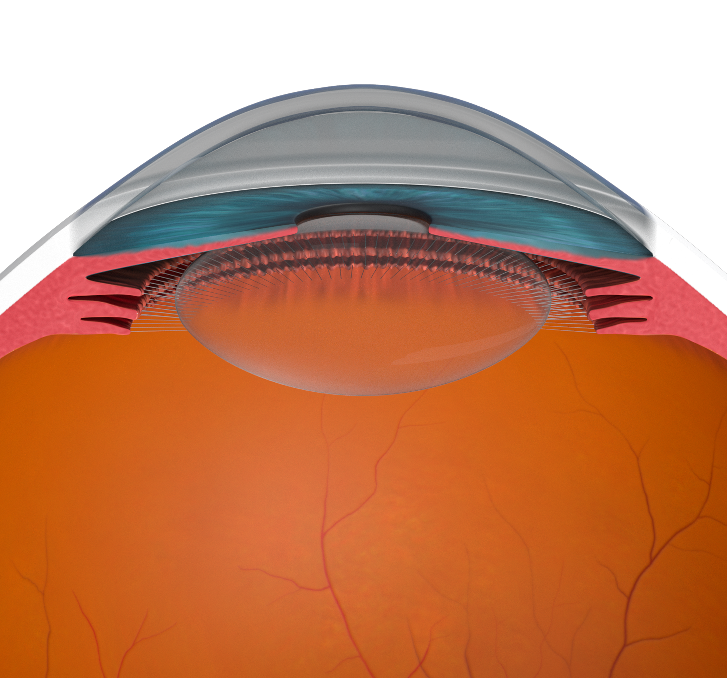
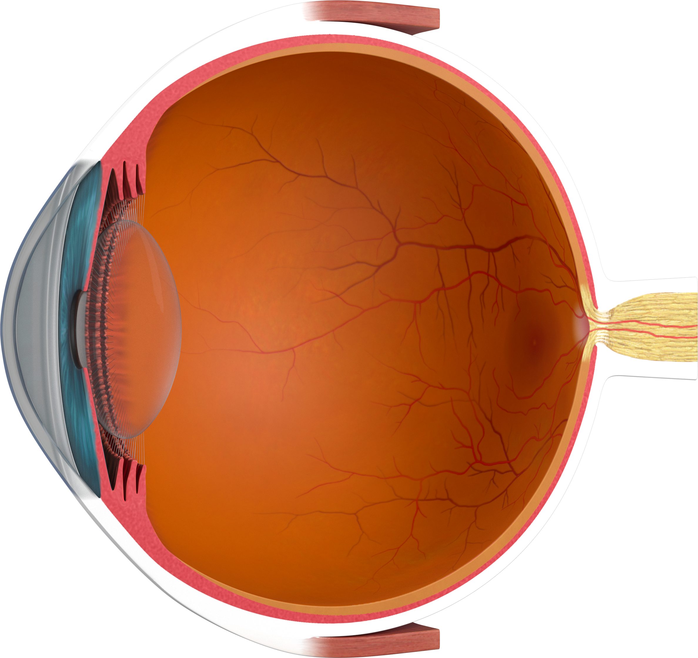
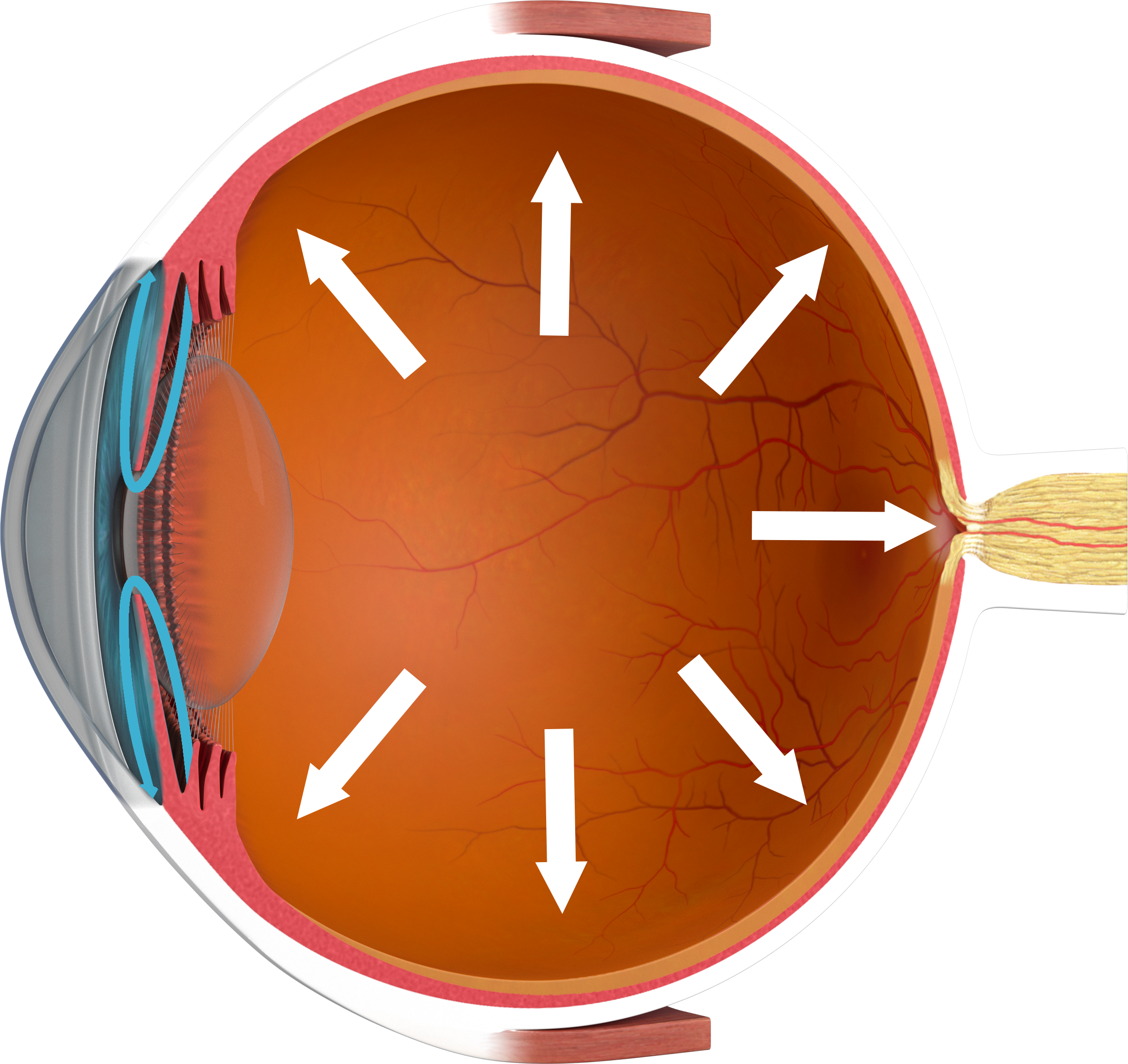
Vision involves the light sensitive photoreceptors cells in the retina absorbing light, a process which naturally creates waste products. Under normal circumstances, these waste products are broken down by the retinal pigment epithelium (RPE) and transported away from the eye by the blood supply. This process becomes less efficient with age.

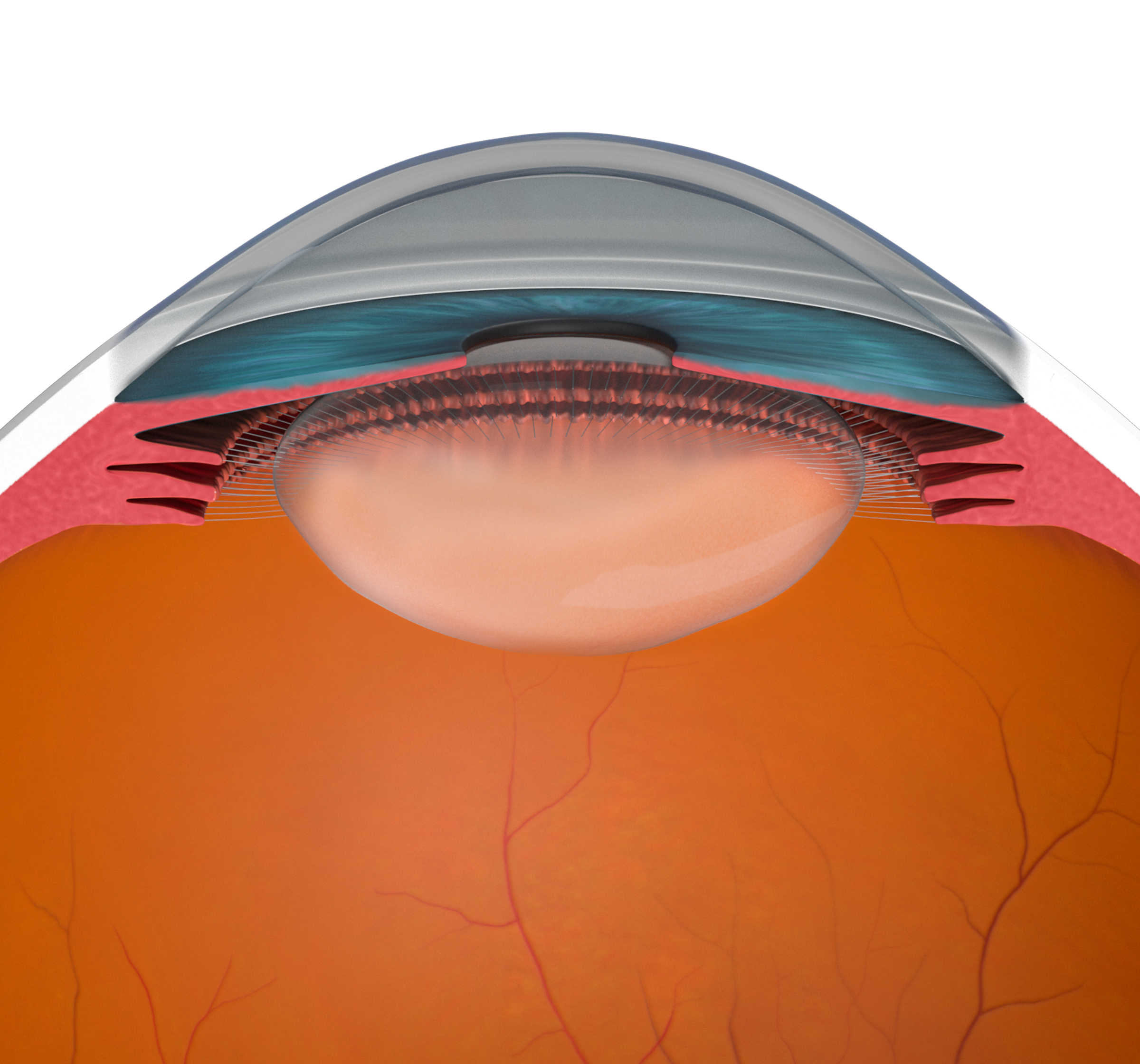
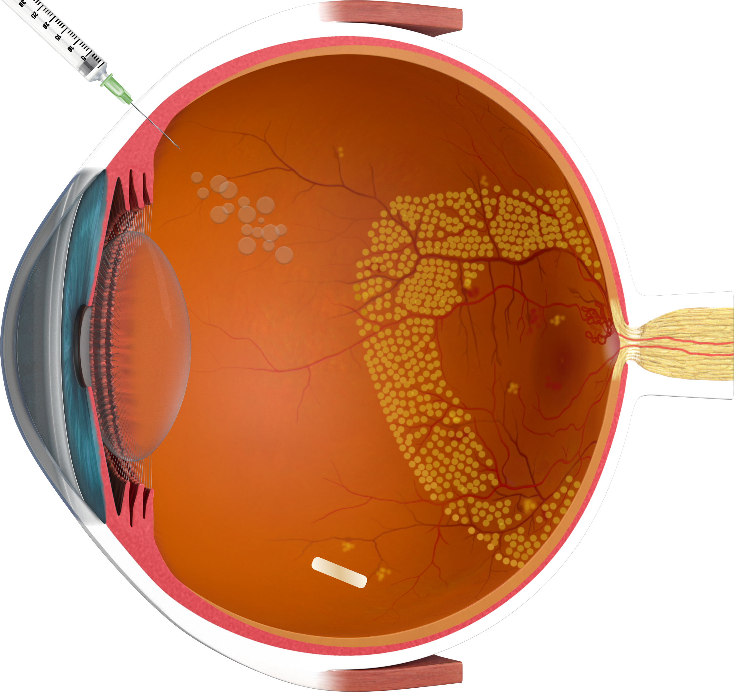
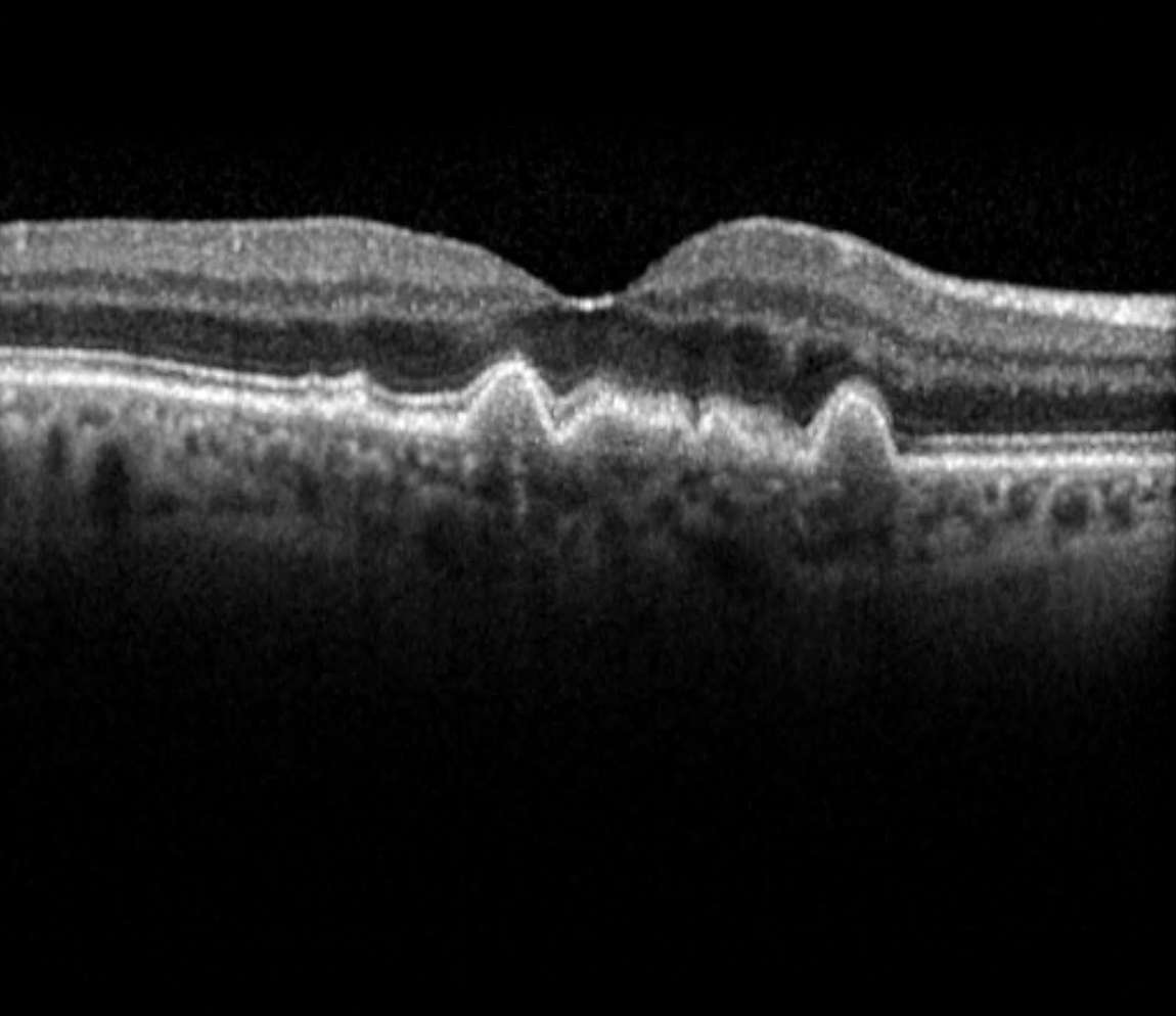
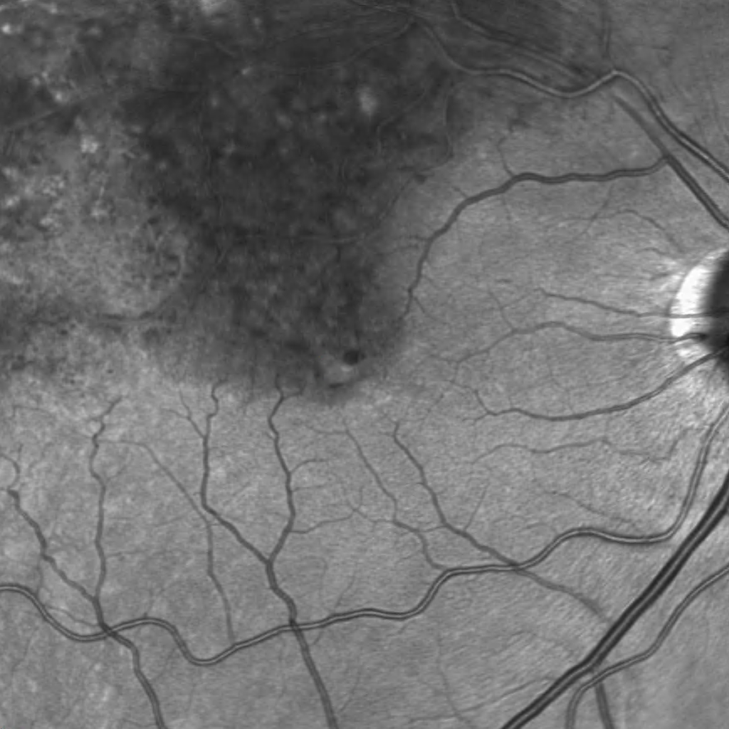
In dry AMD, these degraded products cannot be removed sufficiently, leading to a gradual build-up of waste products, so-called drusen, beneath the RPE cell layer. If the number and size of the drusen increase significantly over time, some RPE cells become damaged and may die. The photoreceptor cells above can no longer be adequately supplied with nutrients and can also die, leading to vision loss in this area. In its late stage, dry AMD is referred to as Geographic Atrophy.

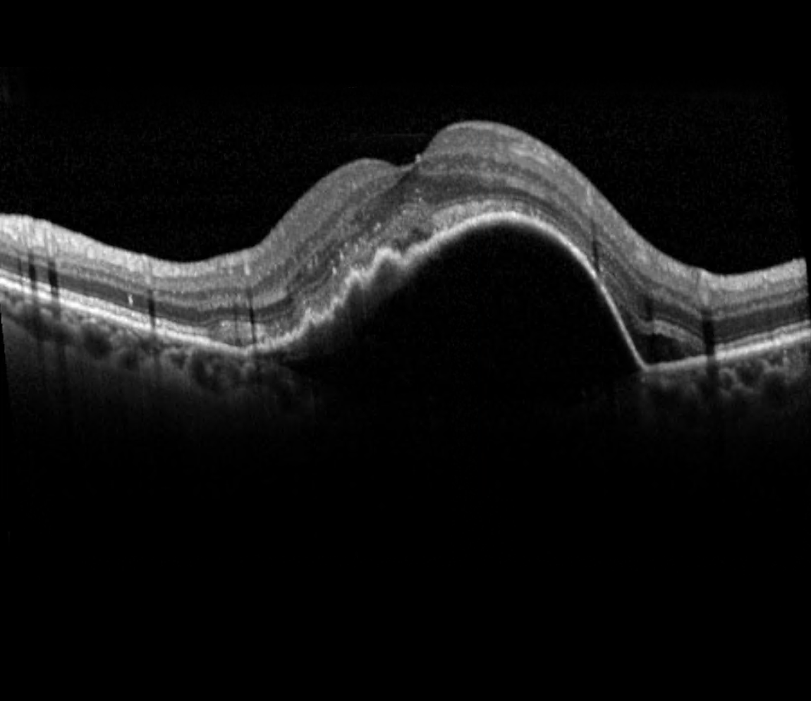
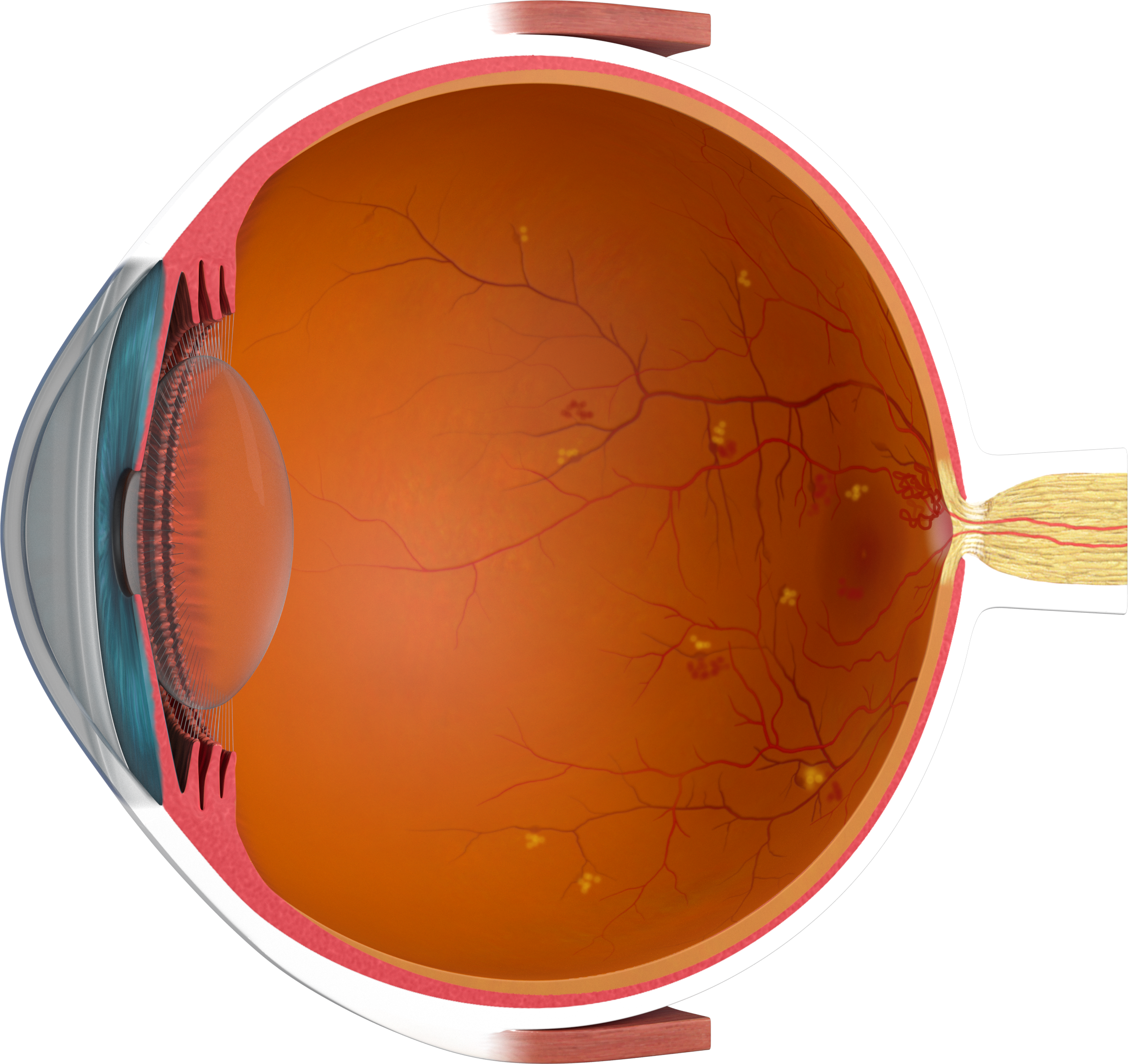
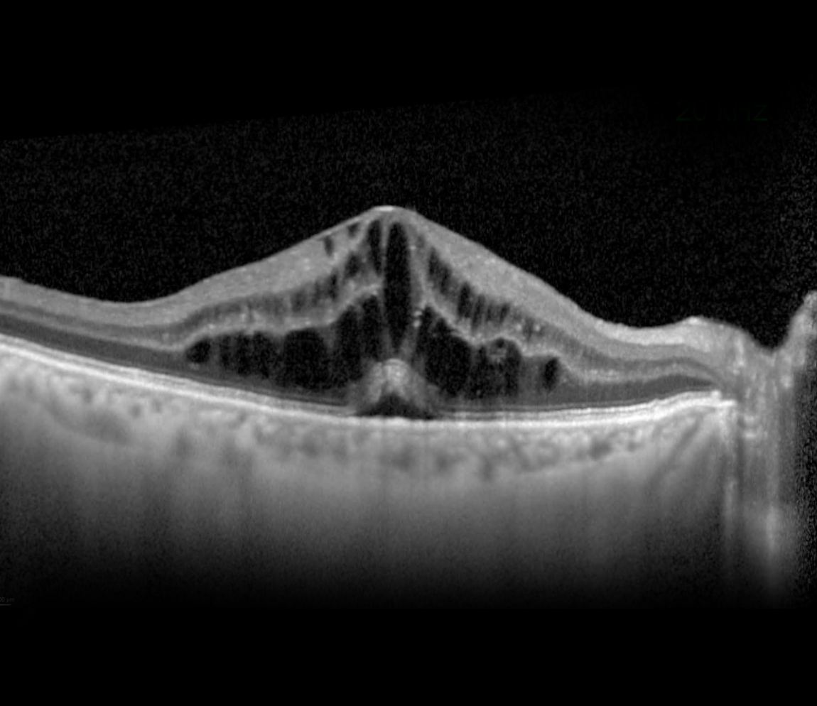
In wet AMD, the build-up of drusen and degeneration of the RPE has started to impair the supply of oxygen to the photoreceptor cells. In response, the retina releases the signaling molecule VEGF, to encourage the formation of new, blood vessels within the retina. However, such new vessels are structurally immature with fragile vessel walls that can suddenly leak fluid/blood. The fluid forms little cystoid spaces within or beneath the retina, distorting vision. Left untreated, permanent vision loss may occur. 

The risk of AMD increases with age, especially after 70, and is influenced by family history and unhealthy lifestyle factors such as smoking, poor diet, physical inactivity, high blood pressure, obesity, and diabetes.

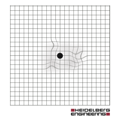
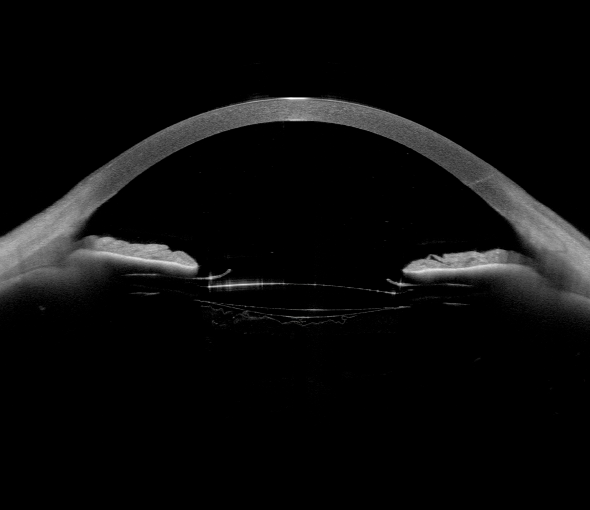
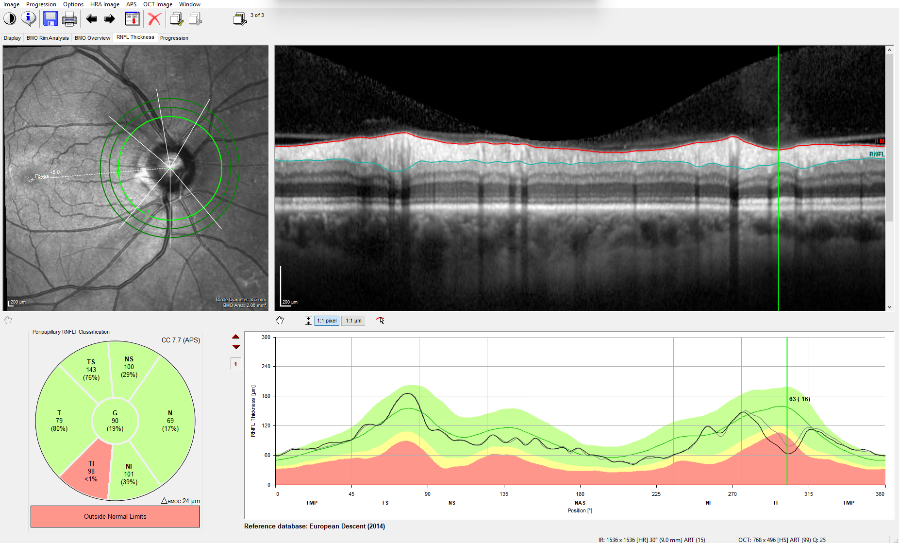
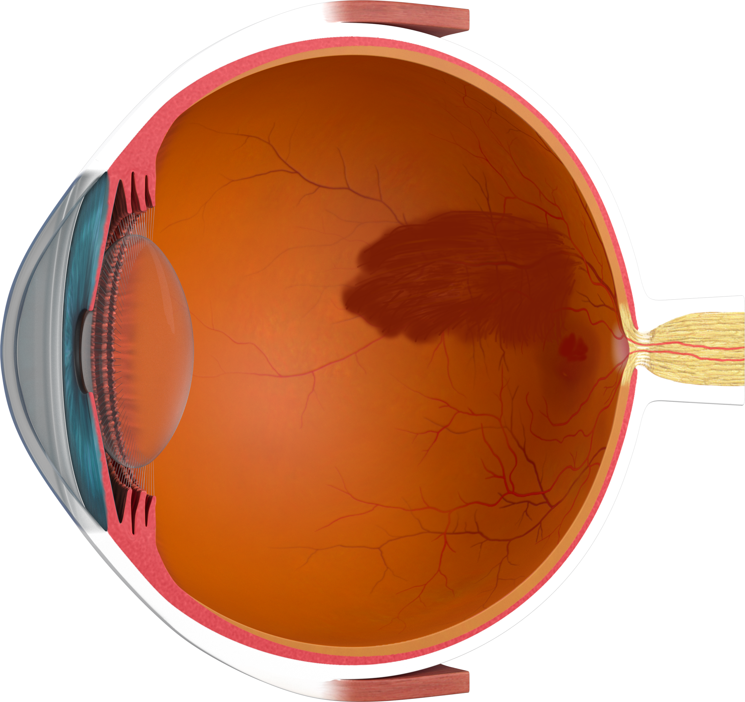
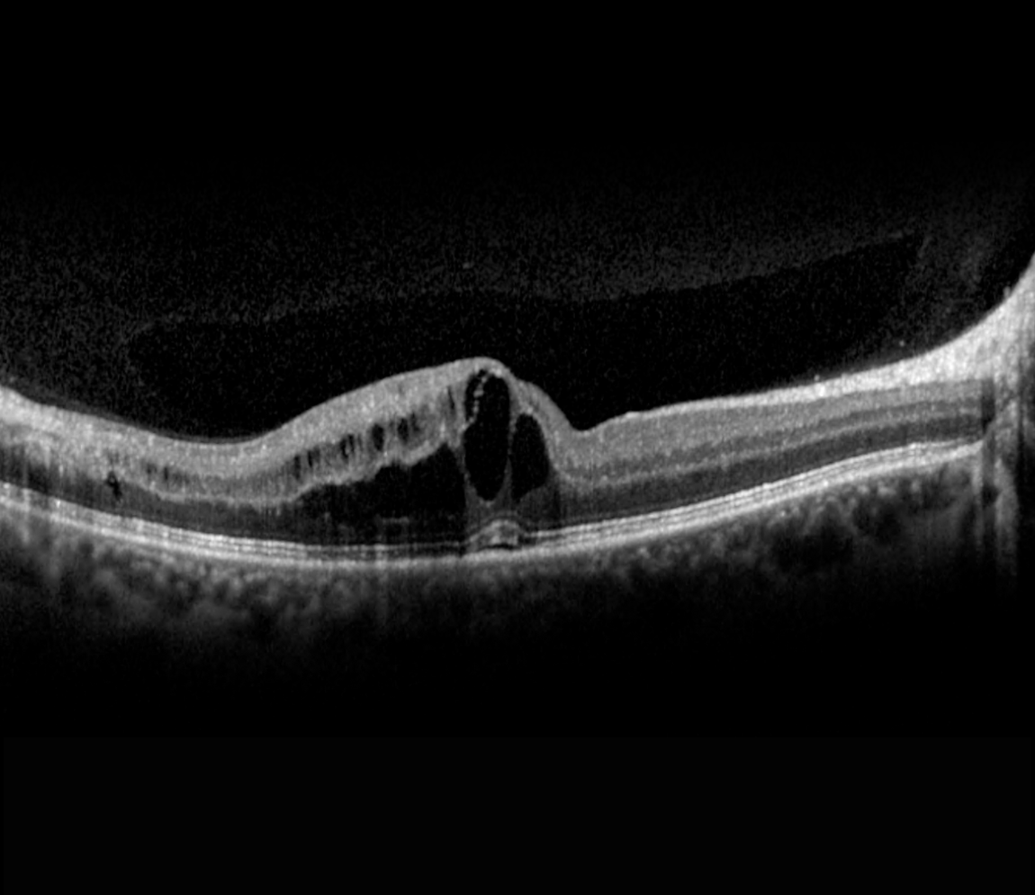
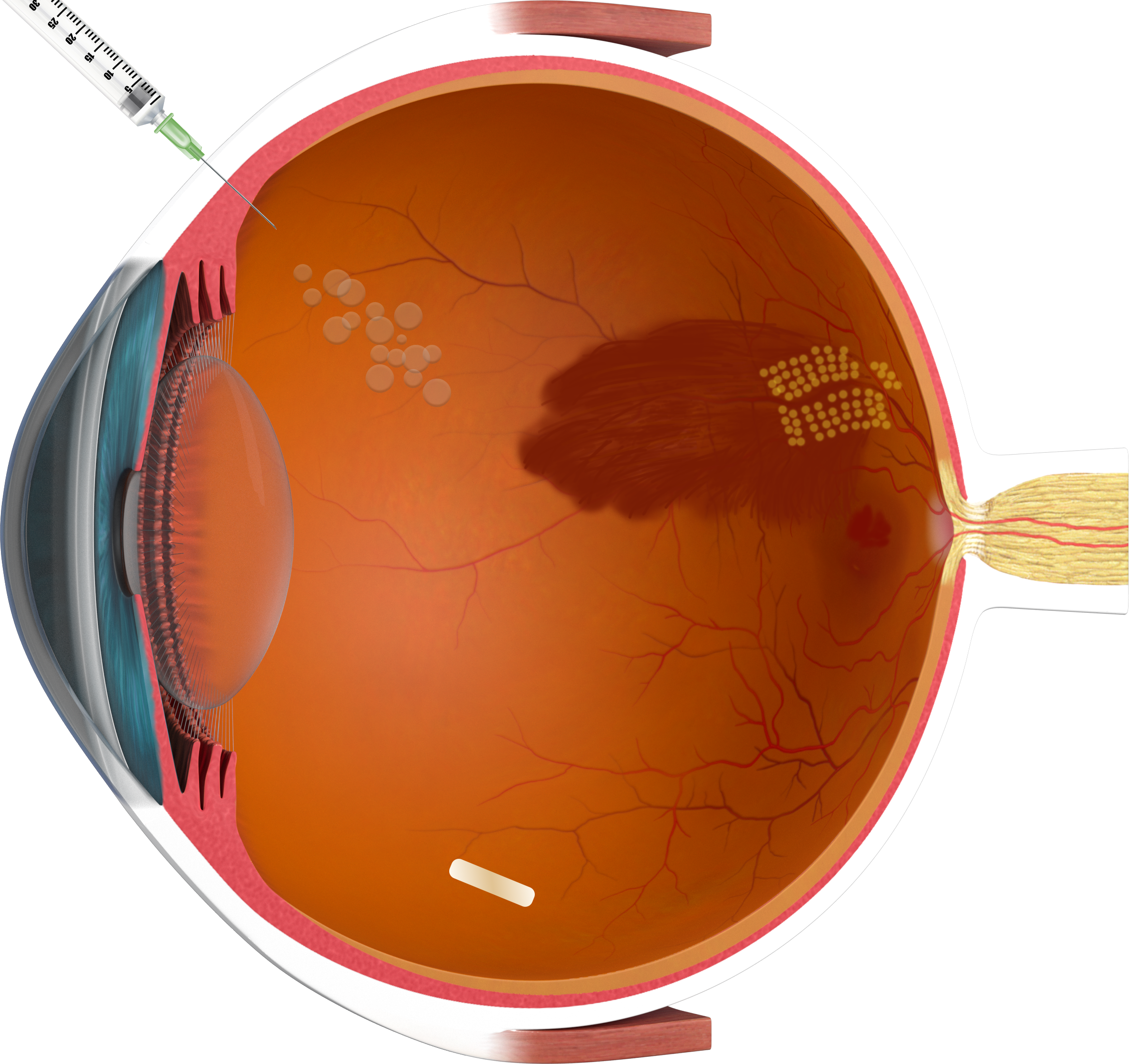
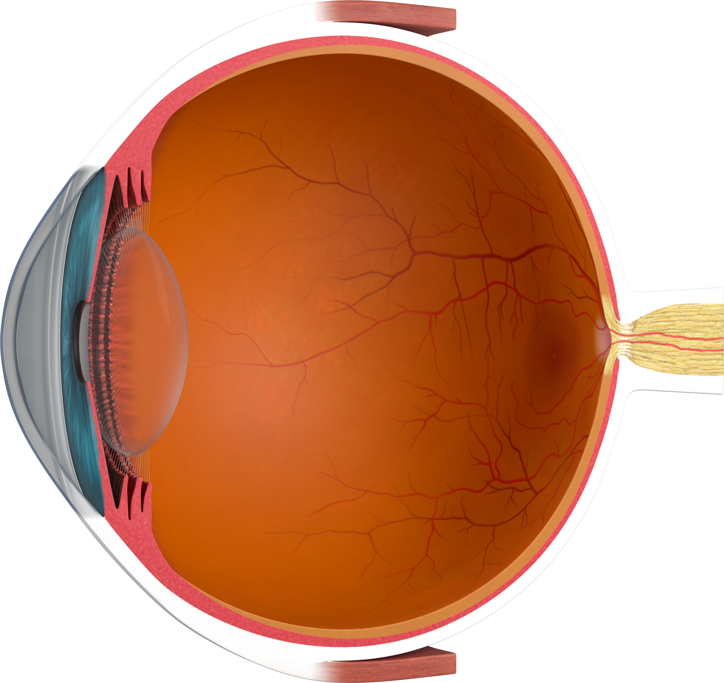
The image illustrates changes in the macula such as cystoid changes and drusen.

Cataract

Cataract is one of the most common causes of treatable vision loss worldwide, especially in people over the age of 60. The crystalline lens of the eye is naturally clear and is responsible for focusing incoming light rays to produce a sharp image on the retina. In cataract, the lens of the eye gradually becomes cloudy, most commonly due to aging, causing incoming light to be scattered or blocked, resulting in poor vision.

Anything causing significant changes to the structure of the crystalline lens, reduces its transparency. This can be an injury to the eye, swelling of the lens related to poorly controlled blood sugar levels in diabetes, but most, simply due to the natural aging process.

Throughout life, the crystalline lens develops more lens fibers, causing it to thicken and become denser and harder. This process gradually reduces the ability for the lens to change shape to focus at different distances.

In addition, the lens fibers degrade and the water content decreases, leading to clouding of the eye lens.

The image illustrates the clouding and thickening of the eye lens in cataracts.

Diabetic Retinopathy (DR)

Diabetes causes increased levels of sugar in the blood, known as hyperglycemia. Poorly controlled blood sugar levels over a long period of time, cause damage to the smallest blood vessels around the body. When the tiny blood vessels in the retina are damaged, this is known as diabetic retinopathy.

Non-proliferative DR
A persistently elevated blood sugar level in diabetes damages structures in the walls of the tiniest blood vessels in the retina—the retinal capillaries. As a result, the vessel walls become unstable, form small pockets called microaneurysms, and turn permeable to lipids (hard exudates), fluid, and/or blood. When fluid builds-up in the macular region of the retina, this is referred to as diabetic macular edema. The damage to the blood vessels also disrupts the oxygen supply to the retina, leading to retinal ischemia, causing permanent damage to the retinal cells.

Proliferative DR
With increasing oxygen deprivation of the retina, the retina attempts to compensate by forming new blood vessels (neovascularization) that tend to grow toward the vitreous body. These newly formed vessels are fragile and can bleed (vitreous hemorrhage) and cause scarring that may result in retinal detachment.

The image illustrates retinal changes in diabetic retinopathy—hard exudates, microaneurysms and intraretinal hemorrhages.


Glaucoma

Glaucoma is the second leading cause of blindness worldwide. It results from damage to the nerve fibers within the retina and optic nerve, commonly as a result of intraocular pressure (IOP) that is too high for an individual eye. If left untreated, this damage can lead to permanent vision loss.  

In open-angle glaucoma the anterior chamber angle is open, but the drainage of aqueous humor through the trabecular meshwork is obstructed, causing an increase in intraocular pressure (IOP > 21 mmHg). 
The cause of normal-tension glaucoma (IOP within the normal range ≈ 10–21 mmHg) has not yet been clearly established. Impaired blood flow, genetic factors, and other mechanisms are under discussion.

In narrow-angle glaucoma or acute closed-angle glaucoma (case of emergency), the anterior chamber angle is too narrow or blocked, which impedes the drainage of aqueous humor and leads to a rapid increase in pressure. 

Risk factors for developing glaucoma include advanced age, family history, ethnic origin and high myopia

The image illustrates a blockage in the drainage of aqueous humor, which results in increased intraocular pressure and damage to the retinal nerve fiber layer.


Retinal Vascular Occlusion

A retinal vascular occlusion occurs when a blood vessel in the retina becomes blocked, disrupting the blood supply to the retina at the back of the eye. This can cause sudden changes in vision and, in some cases, permanent vision problems.

Retinal artery occlusions occur when blood flow in a retinal artery is suddenly blocked. Common causes are emboli (small blood clots or deposits) that originate in other parts of the body—such as the carotid arteries or the heart—and travel into the small blood vessels of the retina. Severe hardening and narrowing of the arteries (atherosclerosis) can also reduce or completely block blood flow.
Major risk factors include high blood pressure, diabetes, elevated cholesterol levels, heart rhythm disorders, and smoking. 

Retinal vein occlusions develop due to impaired blood outflow from the retina. At points where arteries cross over veins, a vein can be compressed by a thickened, hardened artery, leading to blood congestion and eventually to a blockage of the vein.
Contributing factors include high blood pressure, diabetes, elevated blood lipid levels, and blood clotting disorders. Smoking and older age also increase the risk. 

Depending on where the blockage occurs, vascular occlusions are classified as central or branch retinal artery or vein occlusions.

The image illustrates changes in branch retinal vein occlusions.  

Uncover our Products