Acanthamoeba Keratitis
Acanthamoeba can cause several infections in humans, including keratitis, a local infection of the cornea of the eye. This is usually accompanied by severe pain and can result in permanent visual impairment or blindness.
Acanthamoeba can cause several infections in humans, including keratitis, a local infection of the cornea of the eye. This is usually accompanied by severe pain and can result in permanent visual impairment or blindness.
Microscopic, free-living protozoa that are relatively common in the natural environment. Although they can cause infections, most people are exposed to acanthamoebae during their lives and do not contract the disease.
In Ophthalmology, accommodation describes the ability of the eye to change its focus from distant to near objects and vice versa. This process is achieved by the lens changing its shape due to muscle contraction.
A disorder of visual perception that develops during childhood.
A test for the early detection of near vision problems, specifically metamorphopsia and central visual field loss (scotoma).
The space anterior to the iris and crystalline lens.
Angle formed between the back surface of the cornea and the front surface of the iris where they meet.
In anti-VEGF treatment, VEGF inhibitors are injected directly into the vitreous body of the eye. These drugs block the vascular endothelial growth factor (VEGF), which reduces the growth of abnormal blood vessels.
This fluid is produced in the ciliary body. From there it passes through the pupil into the anterior chamber. It consists of water, electrolytes, protein, sugar, ascorbic and hyaluronic acid. It supplies nutrients to many internal eye structures and its production and outflow is responsible for maintaining intraocular pressure.
Astigmatism is most commonly caused by an irregularly shaped cornea, and in rare cases by the lens of the eye. Vision can be impaired, as the eye does not focus light evenly on the retina. Objects appear stretched and distorted. Mild astigmatism does not affect vision and does not require treatment.
Severe hardening and narrowing of the arteries.
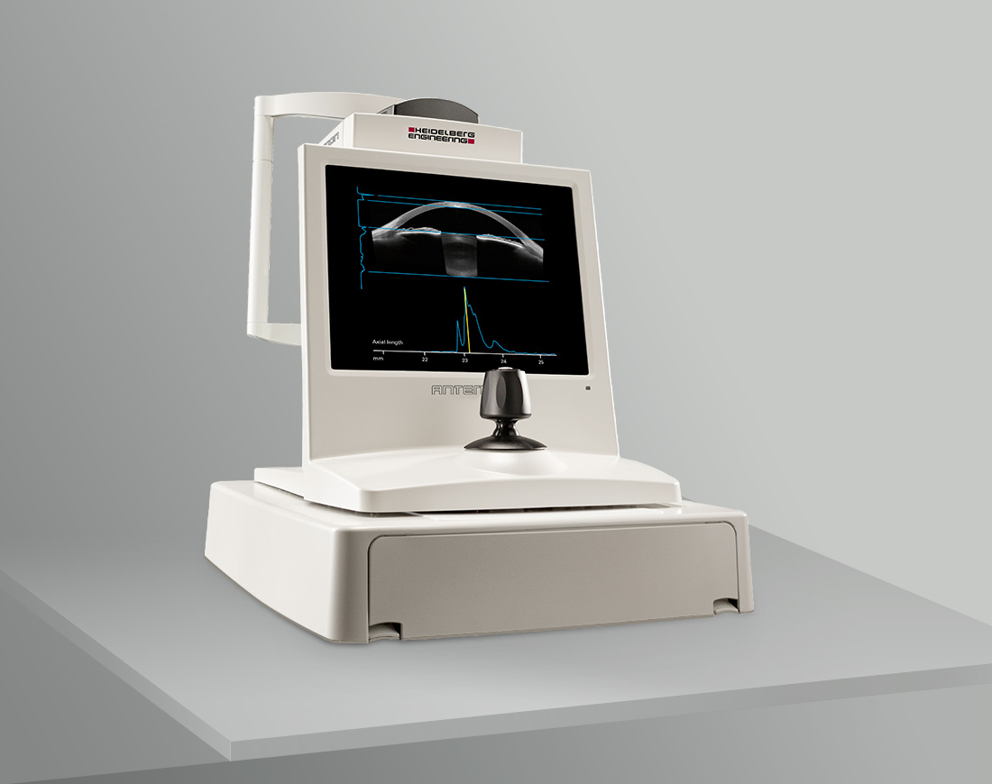
Measurement of the length of the eyeball, and certain distances and structures within it.
Complete or partial vision loss. Blindness is a very severe impairment of visual acuity and/or visual field. It is defined by law in every country. In Germany, among other things, blindness is defined as having a visual field of less than 5° or a visual acuity of the better corrected eye of no more than 0.02, which roughly corresponds to 2% vision. In the UK, blindness is defined as having a visual acuity of less than 3/60 in the better corrected eye, which roughly corresponds to 0.05 % vision. In the US, someone is legally blind when, among other things, having a visual field of less than 20° or a visual acuity of less than 20/200 in the better corrected eye, which roughly corresponds to 1 % vision.
Vascular layer of the eye between the retina and the sclera, providing oxygen and nutrients to the outer retina.
Ring-shaped structure in the eye that produces aqueous humor and controls the shape of the lens for focusing (accommodation).
It lines the space between the eyelids, the eyeball, and the eye socket.
The ability to perceive differences in contrast and distinguish objects from their surroundings.
Hormones produced naturally by the body. When produced artificially, they are used as medications to reduce inflammation in the body.
Located in the posterior chamber. With the refractive power of the cornea (approximately 43 D) the crystalline lens provides additional refractive power of approximately 20 D for accurate focusing of images onto the retina.
Laser procedure that directly targets the ciliary body to reduce its production of aqueous humor, thereby lowering intraocular pressure.
An accumulation of lipids and proteins beneath the RPE.
An accumulation of fluid in the tissue that causes swelling.
Particles that partially or completely block a blood vessel.
Flat cells that coat the inside of blood vessels and help stop the leakage of fluid out of the vessel into the surrounding tissue.
Imaging examination technique in which a fluorescent dye is injected to visualize and assess the perfusion of blood in the retina and choroid of the eye.
A small pit in the retina of the eye where visual acuity is highest (yellow spot or macula). The center of the field of vision is focused in this region where a high number of photoreceptor cells are located.
The back of the eye with its anatomical structures.
A non-invasive imaging technique for assessing retinal health. It detects the natural fluorescence of lipofuscin in the retinal pigment epithelium (RPE), which becomes visible under blue and green laser light. Lipofuscin accumulates as a waste product over the course of a lifetime.
An imaging method that photographs the back of the eye.
Radial light phenomenon around light sources that impairs vision.
Deposits of lipids in the retina that develop when small lipid particles leak from the blood vessels and accumulate in the tissue as a result of persistent fluid retention.
High blood sugar level. It can be caused by various factors and diseases, but diabetes is one of the most common.
An artificial lens that can be implanted into the eye.
The pressure inside the eye that maintains the internal stability of the eyeball.
Into the vitreous body, for example, an intravitreal injection.
Minimally invasive laser procedure in which a tiny opening is made in the iris using a laser to reduce intraocular pressure.
Separates the anterior chamber and posterior chamber. It regulates the amount of light entering the pupil. Two iris muscles control the iris shape and diameter.
Reduced or absent blood flow, which can lead to oxygen deficiency, functional disorders, and, if left untreated for a longer period of time, tissue death.
An inflammation of the cornea. It can have a variety of causes, including bacteria, viruses and fungi. Keratitis is frequently associated with long-term contact lens wear.
Occurs when the cornea thins and gradually bulges outward into a cone-like shape. Keratoconus progresses painlessly and causes distorted vision and deterioration of vision.
Measurement of corneal curvature and refractive power.
Refractive surgery procedure with the aim to correct myopia, hyperopia or astigmatism by changing the corneal curvature. In comparison to LASIK no flap is created. Instead, parts of the epithelium are pushed to the side, followed by reshaping the cornea using an excimer laser that removes corneal tissue.
Refractive surgery procedure with the aim to correct myopia, hyperopia or astigmatism by changing the corneal curvature. After creating a corneal flap the cornea is reshaped using an excimer laser that removes corneal tissue.
The central area of the retina, which contains the area of sharpest vision.
Distorted vision that causes straight lines to appear curved or blurred.
Tiny pockets of enlargement of the fine blood vessels (capillaries) in the retina, from which fluid or blood can leak. They often appear as the first sign of diabetic retinopathy.
A group of low-impact glaucoma surgeries that use very small incisions and implants to reduce intraocular pressure.
A visual impairment, also known as near-sightedness. The refractive power of the cornea and/or lens is too strong in relation to the axial length. The eye is too long or the refractive power of the whole refractive system of the eye is too high. As a result the focal point is in front of the retina and this causes blurred distant vision.
The growth of new, unstable blood vessels where they don't naturally occur, often caused by a lack of oxygen or inflammation.
Axons of the nerve cells, that are long, tube-like extensions that bundle together on their way to the optic nerve head and transmit electrical nerve impulses to the brain.
Examination method based on optical coherence tomography (OCT) that creates a non-invasive three-dimensional representation of the vascular structures of the retina and choroid without the use of dye.
Examination of the back of the eye using an optical instrument called ophthalmoscope.
In a healthy eye, approximately 1 million nerve fibers combine to the optic nerve, which transmits the converted light signals to the brain.
The point of exit of the nerve fiber bundles, located at the back of the eye.
Non-invasive imaging technique for individual tissue structures using laser light.
Measurement of the corneal thickness.
Refers to involvement of almost the entire retina, primarily the peripheral retina, while typically sparing the macula in the center.
Eye surgery technique in which the vitreous chamber is opened via the pars plana and the vitreous body is removed to allow access to the retina. Finally, the vitreous body is replaced with a special fluid or gas.
Cells in the outer wall of blood capillaries and venules that surround the endothelium with their extensions. They are important in maintaining vascular stability, regulating blood flow, and supporting endothelial function.
Measurement of the visual field.
This is a laser treatment method used to seal leaky blood vessels and reduce the number of VEGF.
Light-sensitive cells (rods and cones) of the retina.
Refractive surgery procedure with the aim to correct myopia, hyperopia or astigmatism by changing the corneal curvature. Unlike LASIK, no flap is created here, instead the epithelium is removed completely. This is followed by reshaping the cornea with an excimer laser, which removes corneal tissue. It takes about a week for the epithelium to regenerate.
Describes the space between iris and vitreous where the crystalline lens and the ciliary body are located.
The black aperture in the center of the iris, which is controlled by two iris muscles regulating the diameter of the pupil and thus the amount of light entering the eye.
Located at the back of eye containing several layers including the light-sensitive photoreceptor cells.
Most posterior layer of the retina located between the photoreceptors and the choroid, responsible for the nutrition and maintenance of the visual cycle.
Minimally invasive laser procedure to improve drainage in the eye and thus reduce intraocular pressure.
An eye microscope that ophthalmologists use to examine the eye as standard practice.
An imaging technique that visualizes an object layer by layer.
Measurement of the pressure inside the eye.
An imaging technique that maps the shape of the surface of an object.
Located in the chamber angle and serves as the primary drainage pathway for aqueous humor. After the aqueous humor has entered the anterior chamber through the pupil, it flows through the trabecular meshwork into the venous system.
Surgical procedure in which an artificial drainage pathway is created for the aqueous humor to reduce intraocular pressure.
Vascular endothelial growth factor—a signaling molecule, causing the growth of new, diseased vessels.
Ability to perceive two points separately from a certain distance.
The visual field is the entire area that a person can see when looking straight ahead, including central and peripheral (side) vision. It shows how much of the surroundings can be seen without moving the eyes or head.
Clear, gel-like substance that fills the space between the lens and the retina, helping maintain the eye’s shape and optical properties.