Image Acquisition
PDF Tutorial | 2 pages
This PDF tutorial for SPECTRALIS describes how to acquire images using the MultiColor module.
Get new perspectives with the Heidelberg Engineering Account. Sign up to access exclusive resources and insights.
To make sure you don't miss any news, sign up for our newsletter!
We are committed to providing quick, reliable solutions that support your work and help enable high-quality patient care and research.
Target group: This online course is intended for users of SPECTRALIS optional imaging modules.
Learning objectives: In this course room, you will find a range of e-learning materials designed to help get you started with image acquisition and analysis of the various optional SPECTRALIS imaging modules.
Do you have any questions regarding the learning materials? – Learning materials team
Image Acquisition
PDF Tutorial | 2 pages
This PDF tutorial for SPECTRALIS describes how to acquire images using the MultiColor module.
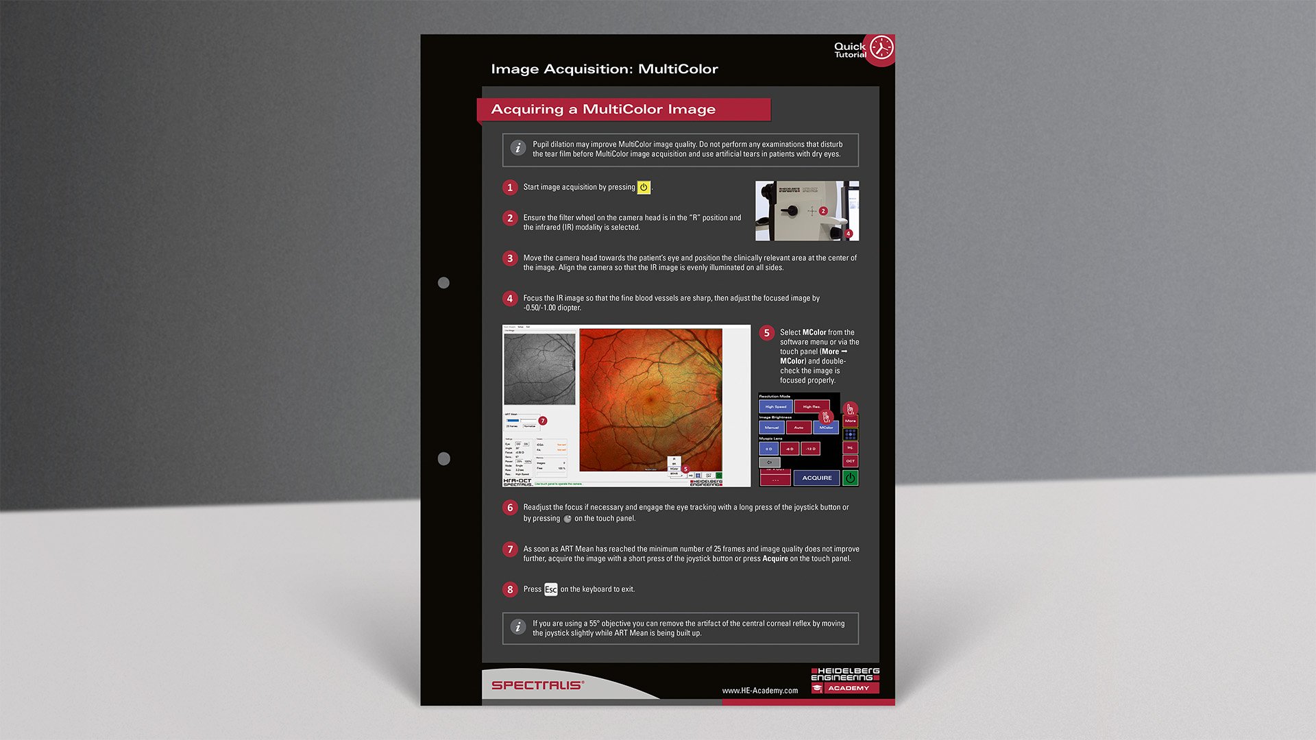
MultiColor Image Acquisition
Video Tutorial | 3 minutes
This video teaches how to acquire MultiColor scanning laser images using the SPECTRALIS imaging platform.
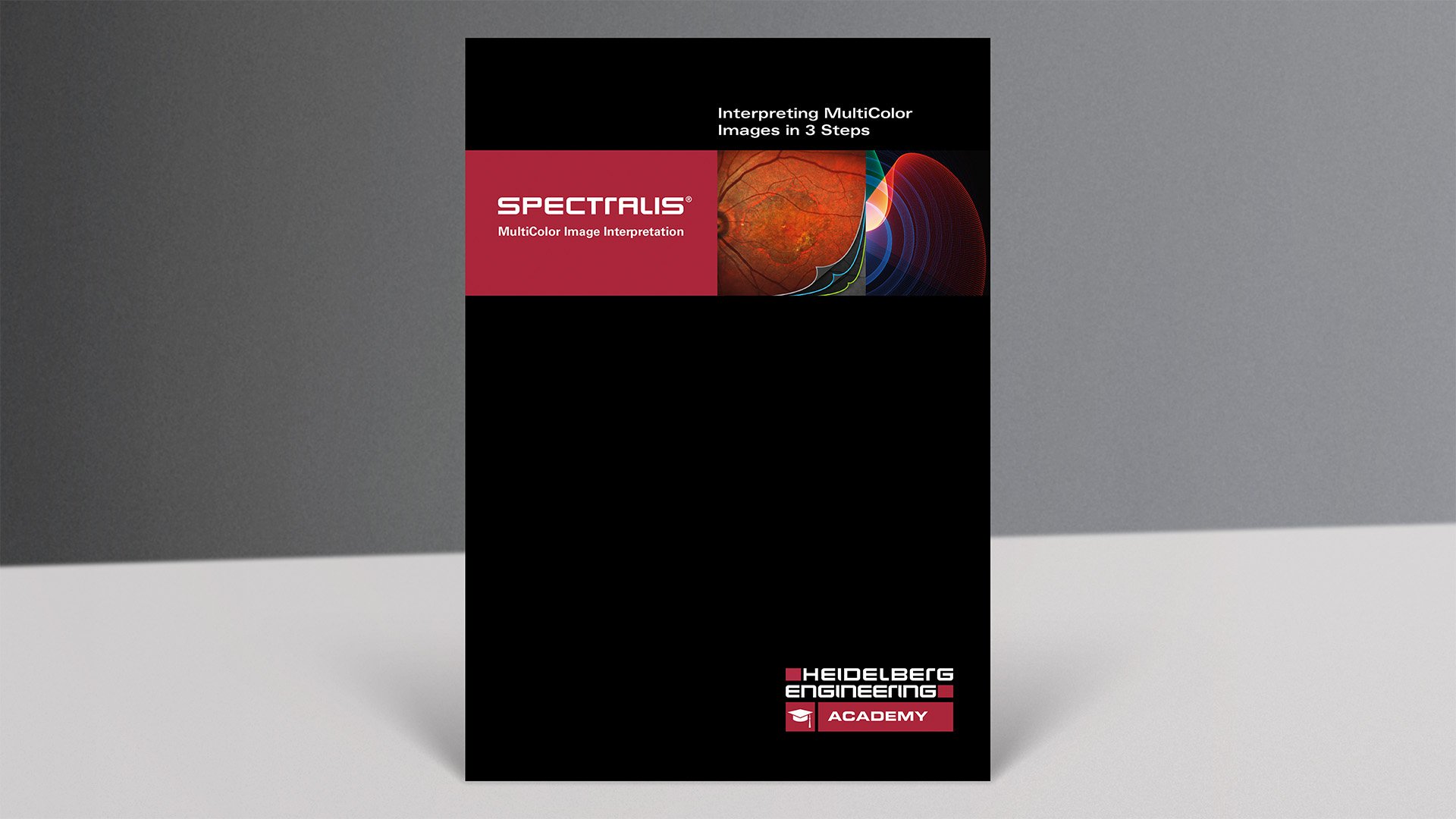
Interpreting MultiColor Images in 3 Steps
PDF Tutorial | 16 pages
This PDF tutorial for SPECTRALIS describes how to acquire images using the MultiColor module.
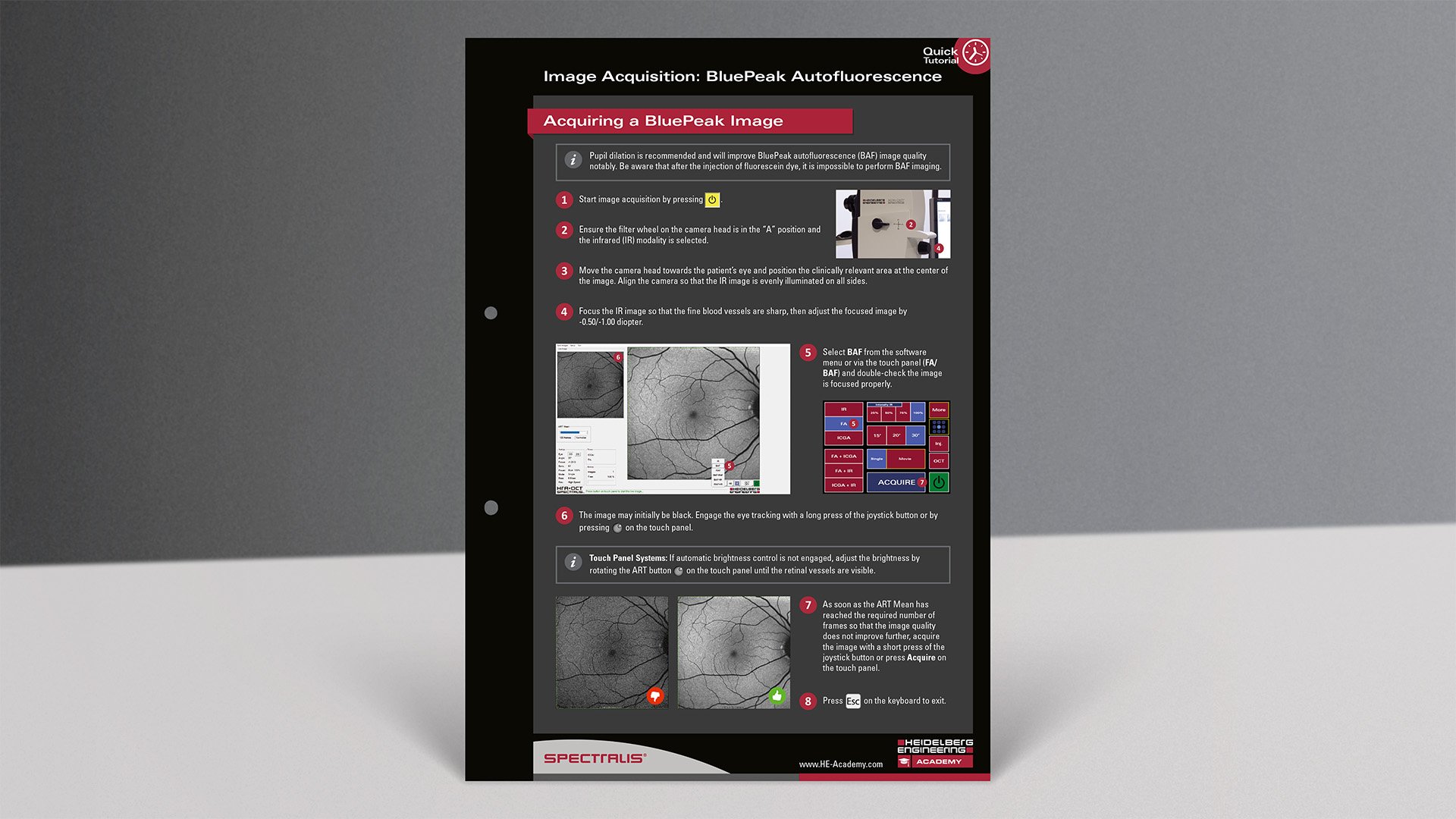
Image Acquisition
PDF Tutorial | 2 pages
This PDF tutorial for SPECTRALIS describes how to acquire images using the BluePeak module.
BluePeak Autofluorescence Image Acquisition
Video Tutorial | 2 minutes
This video teaches how to acquire BluePeak autofluorescence scanning laser fundus images using the SPECTRALIS imaging platform.
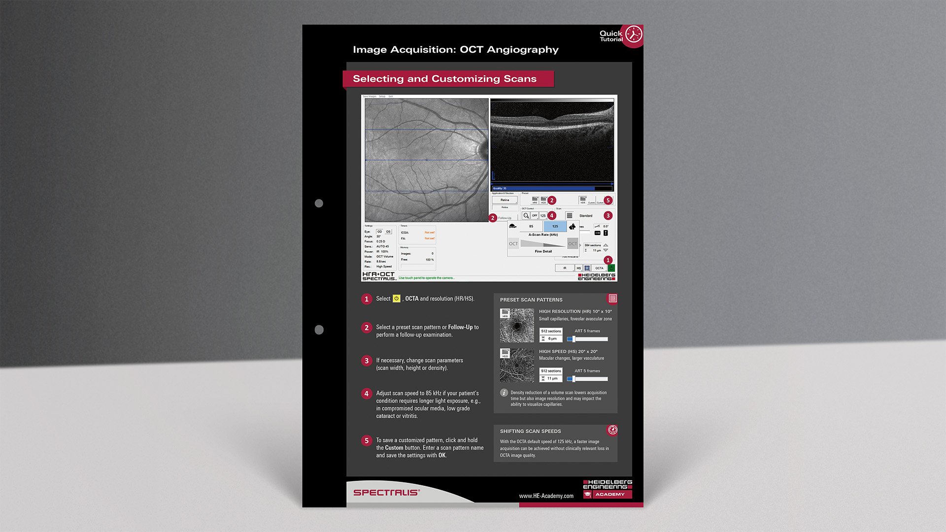
Image Acquisition: OCT Angiography
PDF Tutorial | 2 pages
This PDF tutorial shows you how to customize and perform predefined OCTA scans and check their image quality.
OCT Angiography Module Image Acquisition
Video Tutorial | 8 minutes
This short instructional video provides guidance on how to acquire images using the SPECTRALIS OCT Angiography Module.
Workshop: Angiography Module – Acquisition & Cases
Recorded Webinar | 63 minutes
Learn how to acquire OCT angiography images with SPECTRALIS and to analyze them using tools in the HEYEX software. During the video, Tim Cole, Clinical Affairs Manager, Heidelberg Engineering Limited, demonstrates image acquisition and HEYEX software functionality.
Creating a Custom Slab
PDF Tutorial | 2 pages
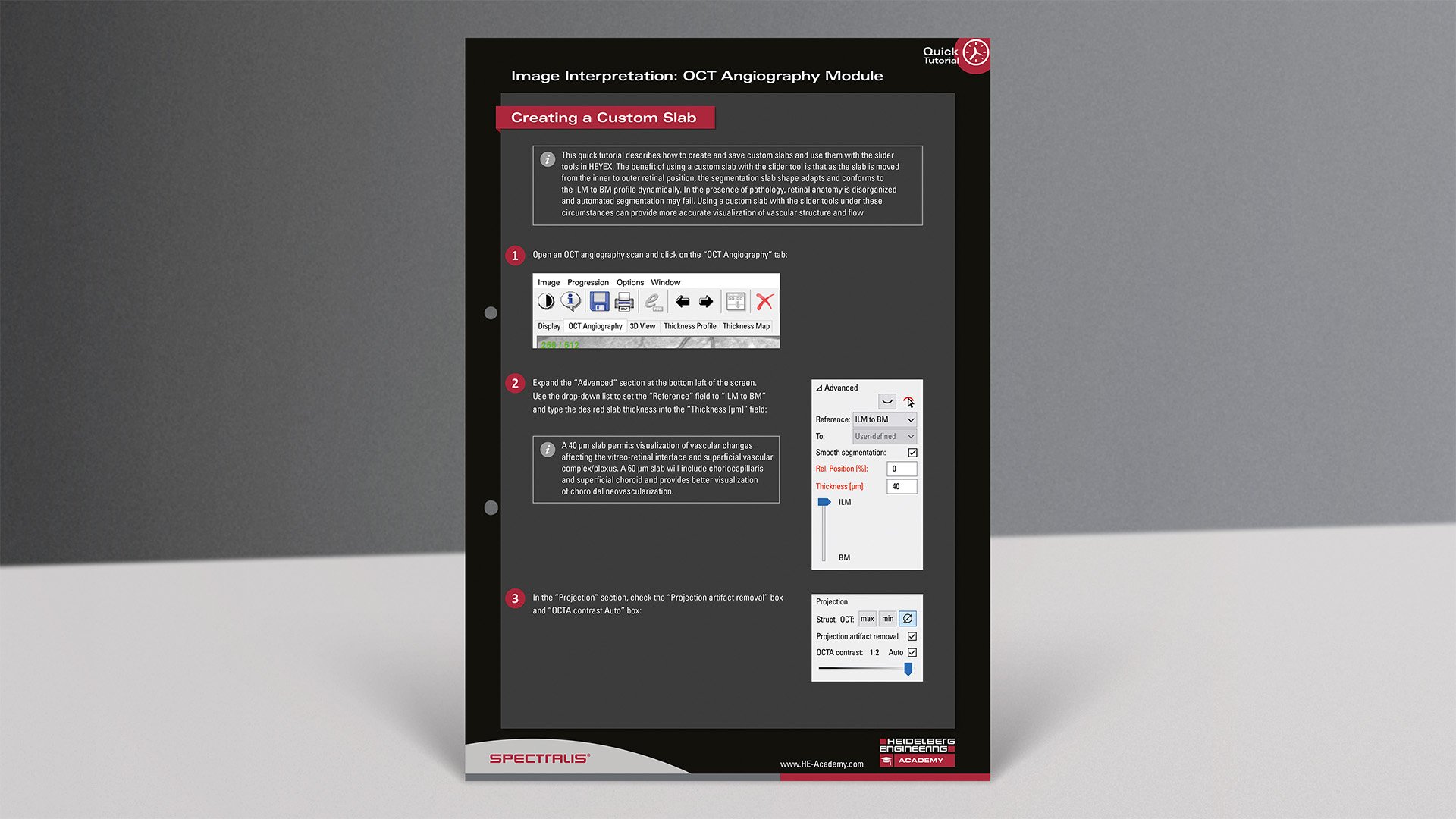
This PDF tutorial describes how to create and save custom OCT angiography slabs and use them with the slider tools in HEYEX.
Systematic Interpretation of OCT Angiography Images
Video Tutorial | 17 minutes
This is a short instructional video which teaches a systematic approach to evaluating OCT Angiography images in 8 steps. The video features Christopher Mody, Director of Clinical Affairs, Heidelberg Engineering Limited and Dr. Richard Gale, Consultant Medical Ophthalmologist, York Teaching Hospital NHS Foundation Trust.
OCT Angiography Imaging & Interpretation: Macular Neovascularization Case Studies
Video Tutorial | 26 minutes
This video tutorial demonstrates the application and benefit of a systematic evaluation of OCT angiography images in a series of patients presenting with various types of choroidal neovascularization (CNV).
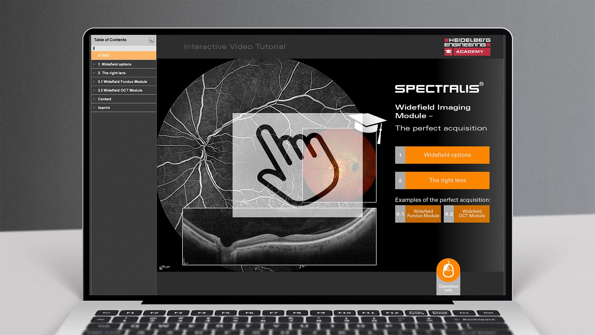
Widefield Imaging Module
Interactive Video Tutorial | 5 minutes
Discover the capabilities of the Widefield Imaging Module and learn how to acquire the perfect image with the new lens!
High Magnification Module Image Acquisition
Video Tutorial | 6 minutes
This short instructional video teaches how to acquire images using the SPECTRALIS High Magnification Module.
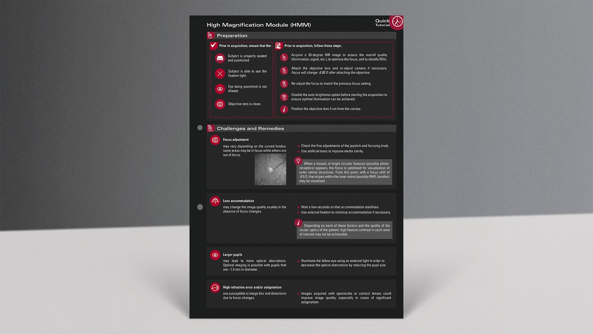
Image Acquisition and Analysis
PDF Tutorial | 2 pages
This PDF tutorial will help you to prepare and perform image acquisitions with the SPECTRALIS High Magnification Module as well as with image analysis and image export.
Workshop: Anterior Segment Imaging
Video Tutorial | 58 minutes
Learn how to acquire multimodal images with the SPECTRALIS anterior segment module and ANTERION and how to analyze them with the tools of the HEYEX software.
Image Acquisition
PDF Tutorial | 6 pages
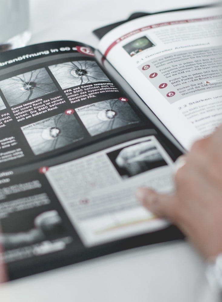
This quick tutorial provides you with practical information about BMO segmentation with the ONH-RC preset for a correct BMO-MRW/RNFLT analysis, troubleshooting within the baseline examination (redefine anatomic landmarks, ΔBMOC > 100 µm) as well as analyzing and monitoring thickness changes within GMPE and creating follow-up reports.
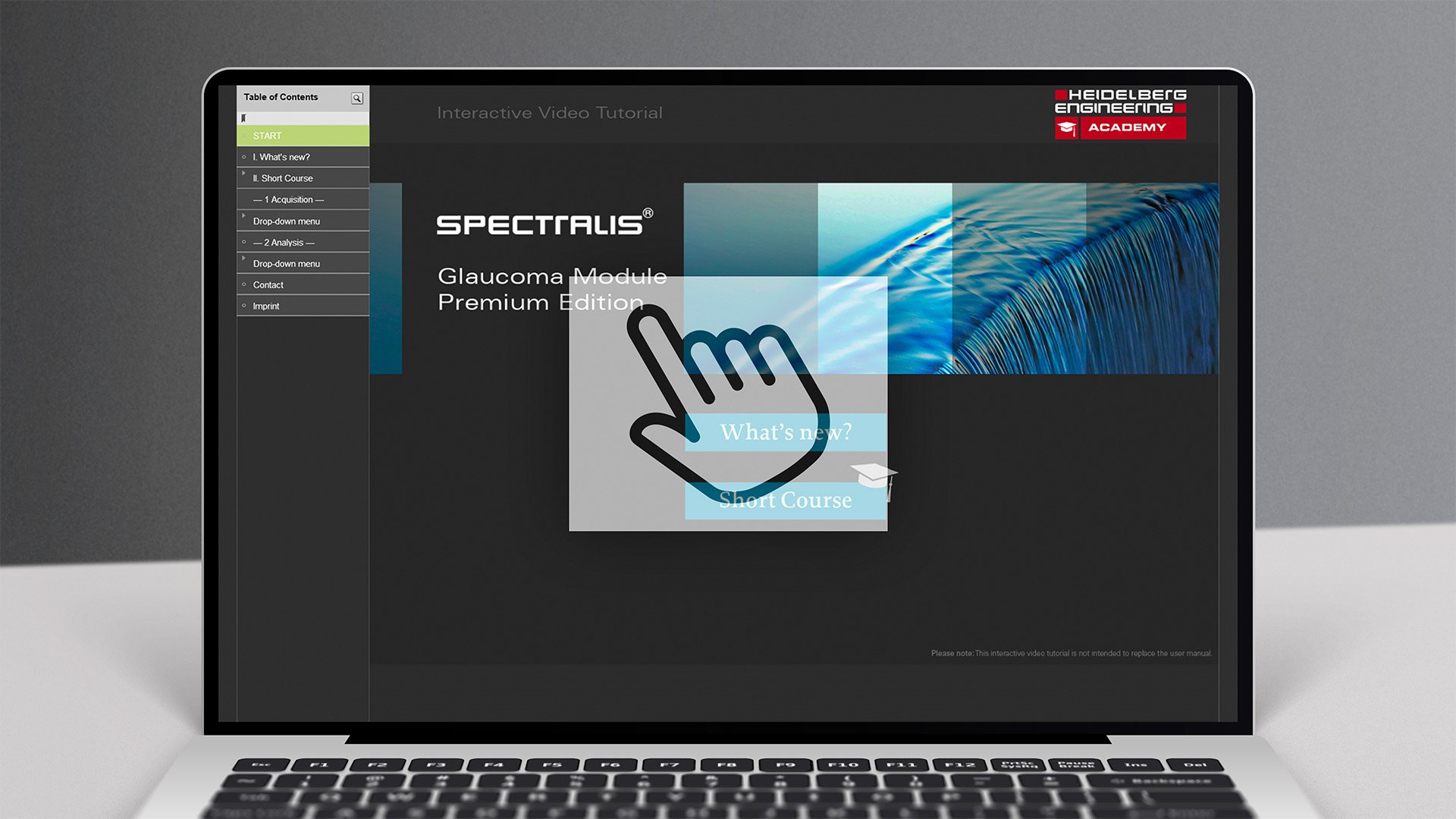
Glaucoma Module Premium Edition
Interactive Video Tutorial | 40 minutes
Get to know the functions of the Glaucoma Module Premium Edition quickly and easily with the help of our interactive video tutorial!
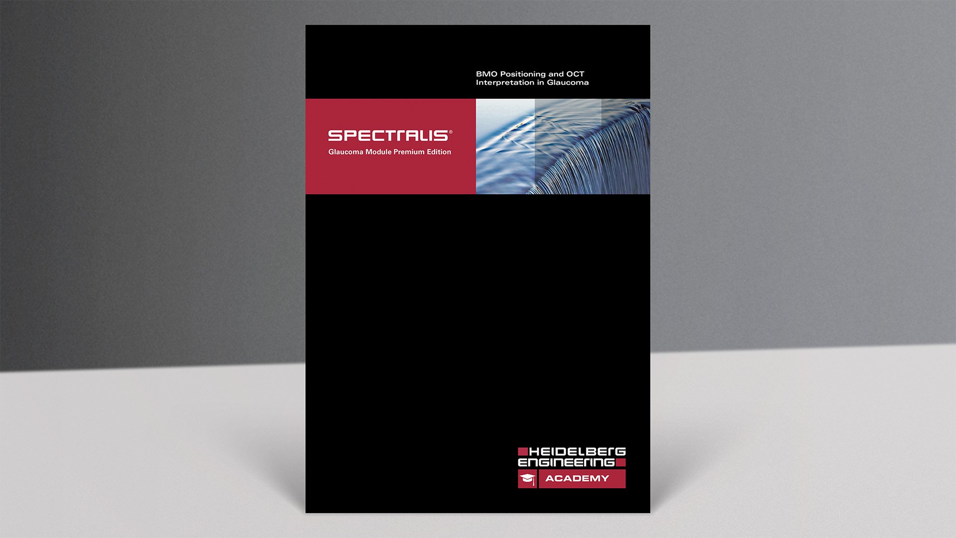
BMO Positioning and OCT Interpretation in Glaucoma
PDF Tutorial | 20 pages
This PDF tutorial contains explanations of the physiological and technical background, the correct positioning of the Bruch's membrane opening (BMO) and the interpretation of the OCT images.
The Glaucoma and Optic Neuropathies Trilogy
Recorded Webinar | 116 minutes
In this recorded webinar, Prof. Dr. Christian Mardin, University Hospital Erlangen, presents the role of OCT imaging in the diagnosis of glaucoma and other optic neuropathies.