POSTERIOR SEGMENT & GLAUCOMA
 High-resolution structural and vascular imaging
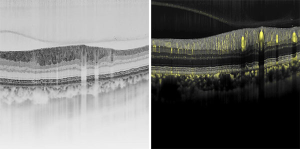
High-resolution structural imaging with OCT is particularly suited for the visualization of chorio-retinal structure, while high-res vasculary imaging with OCT angiography (OCTA) supports researchers in the quest for answers about the connectivity of the vasculature in the eye. Volume renderings of dense B-scan OCTA data have recently been used to characterize macular blood flow connectivity (see link below). Novel high-res OCT and OCTA imaging possibilities could bring researchers and clinicians new insights into the retinal structure and vasculature potentially leading to changes in patient management.
Related Publications
- Atrophic Lesions Associated with Age-Related Macular Degeneration: High-Resolution versus Standard OCT (A. Mahmoudi, G. Corradetti, M. Emamverdi, et al., 2024)
- Cone Density Is Correlated to Outer Segment Length and Retinal Thickness in the Human Foveola (N. Domdei, J. Ameln, A. Gutnikov et al., 2023)
- High-Resolution Optical Coherence Tomography in Healthy Individuals Provides Resolution at the Cellular and Subcellular Levels (J. Reche, A. B Stocker, V. Henchoz et al., 2023)
- Reassessment of hyperautofluorescent rings in retinitis pigmentosa using novel retinal imaging techniques (D. Cabral, P. Ramtohul, K. B. Freund, 2023)
- Macular Vascular Imaging and Connectivity Analysis Using High-Resolution Optical Coherence Tomography (D. Cabral, A. C. Fradinho, Telmo Pereira et al., 2022)
- Deep Capillary Plexus Features in Acute Macular Neuroretinopathy: Novel Insights Based on the Anatomy of Henle Fiber Layer (D. Cabral, P. Ramtohul, L. Zatreanu et al.. 2022)
- Advances in photoreceptor quantification moving from conventional to high-resolution SPECTRALIS optical coherence tomography (Sophie Frank; Hrvoje Bogunovic; Oliver Leingang; Philipp Fuchs; Leonard Coulibaly; Gregor Sebastian Reiter; Ursula Schmidt-Erfurth, 2022)
- Reliability of retinal layer annotation with a novel, high-resolution optical coherence tomography device: a comparative study (Leon von der Emde, Marlene Saßmannshausen, Olivier Morelle,Geena Rennen,Frank G. Holz, Maximilian W. M. Wintergerst, andThomas Ach, 2023)
- Lateral Resolution of a Commercial Optical Coherence Tomography Instrument (R. F. Spaide, T. Otto, S. Caujolle et al., 2022)
- Intermediate and Deep Capillary Plexuses in Machine Learning Segmentation of High-Resolution Optical Coherence Tomography Imaging (R. F. Spaide, T. Otto, S. Caujolle, 2021)
- Volume Rendering of Dense B-Scan Optical Coherence Tomography Angiography to Evaluate the Connectivity of Macular Blood Flow (D. Cabral, T. Pereira, G. Ledesma-Gil et al., 2020)
- Treatment of Sorsby fundus dystrophy with anti-tumor necrosis factor-alpha medication (R. F. Spaide, 2022)
 Time-dependent fluorescence imaging
Fluorescence Lifetime Imaging Ophthalmoscopy (FLIO) is an experimental non-invasive imaging modality that provides another fluorescence dimension. In the past, the intensity or the spectrum of retinal fluorescence has been explored. But with FLIO, researchers are able to observe the time dependency of the intrinsic fluorescence of the retina. This capability adds a new dimension to the images, which yields phenotypic results that may be indicative of alterations in retinal metabolism preceding structural changes. Recent studies show that for many retinal pathologies, such as AMD, Stargardt’s disease, MacTel type 2, as well as in conditions such as chloroquine toxicity, FLIO may show distinct patterns for either better staging of disease progression or even indicating earliest changes.
Related Publications
- Progressive Dysmorphia of Retinal Pigment Epithelium in Age-Related Macular Degeneration Investigated by Fluorescence Lifetime Imaging (M.Hammer, J. Jakob-Girbig, L. Schwanengel et al., 2021)
- Comparing Fluorescence Lifetime Imaging Ophthalmoscopy in Atrophic Areas of Retinal Diseases (L. Goerdt, L. Sauer, A. S. Vitale et al., 2021)
- Longitudinal Foveal Fluorescence Lifetime Characteristics in Geographic Atrophy using Fluorescence Lifetime Imaging Ophthalmoscopy (J.-B. Lincke, C. Dysli, D. Jaggi et al., 2021)
- Fluorescence lifetime imaging ophthalmoscopy (C. Dysli, S. Wolf, M. Y. Berezin et al, 2017)
- Fluorescence lifetime imaging ophthalmoscopy: autofluorescence imaging and beyond (L. Sauer, A. S. Vitale, N. K. Modersitzki et al.,2021)
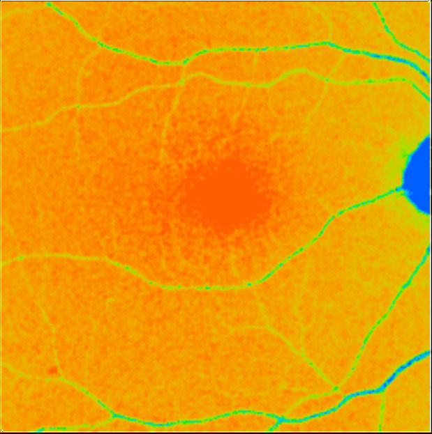
Macular pigment assessment
Standardizing the assessment of macular pigment has been inherently difficult since existing techniques are unable to provide a full image and measurements are subjective and highly variable. Among others, Prof. John Nolan and his group have contributed to the establishment of a comprehensive and objective evaluation of the macular pigment profile. In 2020, he received the Heidelberg Engineering Xtreme Research Award for his efforts to assess the importance of macular pigment for visual performance in both healthy individuals and AMD patients, standardizing these measurements with an investigational SPECTRALIS Macular Pigment Optical Volume Module. This project aimed to link macular carotenoids to cognitive function and brain health, with the goal of identifying ways to reduce the risk of Alzheimer’s disease.
Related Publications
- Comparing 2-dimensional macular pigment optical density with objective and subjective perimetry and visual acuity in age-related macular degeneration (B. B. Rai, F. Sabeti, J. P. van Kleef et al., 2024)
- Macular and Plasma Xanthophylls Are Higher in Age-related Macular Degeneration than in Normal Aging: Alabama Study on Early Age-related Macular Degeneration 2 Baseline (G. McGwin Jr., D. Kar, A. Berlin et al., 2022)
- Deep learning-based correction of cataract-induced influence on macular pigment optical density measurement by autofluorescence spectroscopy (A. Obana, K. Ote, Yuko Gohto et al., 2024)
- Standardizing the Assessment of Macular Pigment Using a Dual-Wavelength Autofluorescence Technique (M. Green-Gomez, P. S. Bernstein, C. A. Curcio et al., 2019)
- Lutein and Zeaxanthin Distribution in the Healthy Macula and Its Association with Various Demographic Factors Examined in Pseudophakic Eyes (A. Obana, Y. Gohto , R. Asaoka et al., 2021)
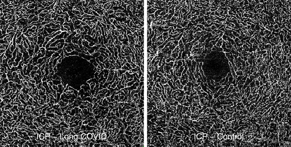
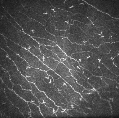
Image courtesy: Mardin, Hohberger, et al.
New insights with OCTA
The Heidelberg Engineering Xtreme Research Award 2022 winner, PD Dr. Dr. Bettina Hohberger, and a research group at the University Clinic in Erlangen, Germany, utilized SPECTRALIS OCTA in an experimental setting to assess therapeutic responses of patients impaired by chronic fatigue syndrome in relation to a preceding COVID-19 infection (“long COVID syndrome”). Long COVID may present certain characteristics in retinal capillary density that can be identified with OCTA.
Related Publications
- Choriocapillaris Impairment Is Associated With Delayed Rod-Mediated Dark Adaptation in Age-Related Macular Degeneration (D. Kar, G. Corradetti, T. A. Swain et al., 2023)
- Confocal MultiColor Signal Depends on Perfusion Characteristics of Retinal Microaneurysms in Diabetic Retinopathy as Detected by OCTA (A. Arrigo, M. Teussink, A. Antropoli et al., 2023)
- Optical coherence tomography angiography macular biomarkers of peripheral retinal ischemia in diabetic macular edema: secondary endpoints from the clinical study “FOVEA” (R. Serra, F. Coscas, J. F. Boulet et al., 2024)
- Reproducibility of peripapillary, optic nerve head and macular vessel density by OCT-A according to glaucoma severity staging (L. Salazar-Quiñones, P. Peña-Urbina, J. I. Fernández-Vigo et al., 2023)
- Retinal Microvasculopathy with Different Insulin Infusion Therapies in Children with Type 1 Diabetes Mellitus without Clinical Diabetic Retinopathy (Y. Guo, X. Zheng, H. He et al., 2023)
- Neutralization of Autoantibodies Targeting G-Protein-Coupled Receptors Improves Capillary Impairment and Fatigue Symptoms After COVID-19 Infection (B. Hohberger, T. Harrer, C. Mardin et al., 2021)
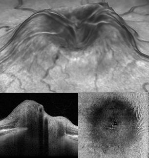 Non-invasive exploration of intracranial hypertension
The winners of the Xtreme Research Award 2021 have brought neuro-ophthalmology to the forefront of ocular imaging and innovation with their “OCT Neuro-Toolbox” approach for the diagnosis and management of papilledema, optic disc edema, and pseudo-papilledema. Their research has pointed to a practical, office-based SD-OCT set of examinations for the distinction of optic disc edema from pseudo-papilledema. OCT exams have so far been largely focused on maculopathies and glaucoma, leading to limitations in the presence of severe edema. Their proposed SD-OCT neuro toolbox is based on the quantitative assessment of mean RNFL and GC-IPL thickness complemented by the qualitative assessment of different SD-OCT imaging options, including an examination of the ONH in adduction to expose potential folds or deformations not evident in primary position.
Related Publications
Selective retina therapy
For the past few years, Heidelberg Engineering has supported a collaborative research project involving several research institutes in Germany and Switzerland focusing on OCT guided selective retinal laser treatment. Selectively removing retinal pigment epithelium (RPE) as a therapeutic intervention may hold potential in the treatment of AMD and other RPE-associated eye diseases. Heidelberg Engineering is providing technology for real-time dosimetry purposes and for visualizing structural information via OCT to support the research. OCT may provide informative feedback on outcomes of selectivity of the treatment.
Related Information
HuCE-optoLab (University of Bern, Switzerland)
Website in German
Related Publications
- Real-time OCT feedback-controlled RPE photodisruption in ex vivo porcine eyes using 8 microsecond laser pulses (C. Burri, S. Salzmann, J. Wandel et al., 2023)
- High-Precision Optical Coherence Tomography Navigated Laser Retinopexy for Retinal Breaks (S. Salzmann, P. Wakili, S. Al-Nawaiseh et al., 2023)
- Investigation of the Influence of Pulse Duration and Application Mode on Microsecond Laser Microsurgery of the Retinal Pigment Epithelium (C. Burri, S. Salzmann, M. Amstutz et al., 2023)
- Selective Large-Area Retinal Pigment Epithelial Removal by Microsecond Laser in Preparation for Cell Therapy (C. Burri, S. Al-Nawaiseh, P. Wakili et al., 2021)
- Optical coherence tomography controlled selective retina therapy with a novel microsecond laser (C. Burri, A. Hutfilz, L. Grimm et al., 2019)
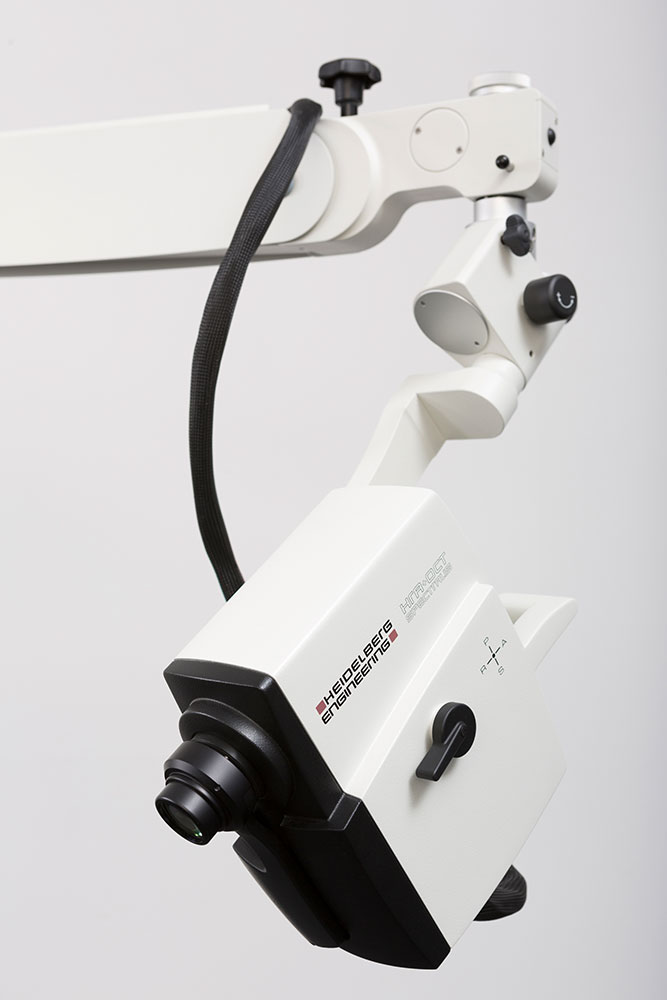 Flexible diagnostic imaging for patients in a supine position
The SPECTRALIS Flex Module*, affixed to a movable stand with an adjustable and highly flexible arm, supports novel research in subjects needing to be examined in a supine position. All SPECTRALIS imaging modalities are available in combination with the Flex Module – without sacrificing image quality.
Related Publications**
- Reduced Aqueous Humor Outflow Pathway Arborization in Childhood Glaucoma Eyes (S. Gupta, X. Zhang, A. Panigrahi et al., 2024)
- Optical coherence tomography (OCT) in unconscious and systemically unwell patients using a mobile OCT device: a pilot study (X. Liu, A. U. Kale, N. Capewell et al., 2020)
- Repeatability and reproducibility of quantitative OCT angiography measurements from table-top and portable Flex Spectralis devices (A. Ponugoti, H. Ngo, S. Stinnett et al., 2024)
- Fluorescein Aqueous Angiography in Live Normal Human Eyes (A. S. Huang, R. C. Penteado, S. K. Saha et al., 2019)
- Aqueous angiography guided ab interno trabecular surgery for open-angle glaucoma (T. Dada, A. N. Bukke. 2021)
- Optical Coherence Tomography Normative Peripapillary Retinal Nerve Fiber Layer and Macular Data in Children 0–5 Years of Age (J. C. Rotruck, R. J. House. S. F. Freedman et al., 2019)
*Not available in all countries
**The applications of the devices or modules as described in the publications are not part of the intended use of the product. The interest of Heidelberg Engineering in these publications is to support scientific research to discover innovative imaging applications to improve patient care.

Please note: We present general research topics and related publications on this page to let science speak for itself. For technologies that are part of our commercial product portfolio, we provide further information and refer to our product web pages. Other technologies showcased are not for sale and are not a reflection of our commercial product releases or planned product developments.


