ANTERIOR SEGMENT
 In vivo cellular imaging
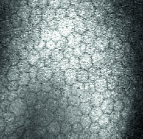
Confocal scanning laser microscopy serves as unique technology to produce high-resolution in vivo images of the cornea and external ocular structures. Corneal microscopes – such as the HRT3 RCM* – make use of this technology, scanning the cornea with a field of view up to 400 x 400 μm. This allows for the documentation of corneal morphology at the cellular level and to identify keratocytes subpopulations. The in vivo assessment of corneal structures can aid in the diagnosis and monitoring of abnormalities, pre- and post-surgery evaluation and in the assessment of dry eye disease.
Related Publications
*Not available in all countries
 High-resolution imaging of nerves and dendritic cells
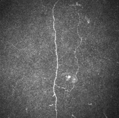
Confocal scanning laser microscopy also serves as a valuable tool to visualize and document the corneal subbasal nerve plexus. High-resolution imaging allows for the investigation and analysis of the fine details of the nerves, which opens a new focus to study nerve structures in diabetic patients as well as patients that are subject to nerve fiber loss resulting from other diseases such as cognitive impairment and dementia.
Related Publications
- Corneal Nerve and Brain Imaging in Mild Cognitive Impairment and Dementia (E. Al-Janahi , G. Ponirakis, H. Al Hamad et al., 2020)
- Association of Cerebral Ischemia With Corneal Nerve Loss and Brain Atrophy in MCI and Dementia (G. Ponirakis, A. Elsotouhy, H. Al Hamad et al., 2021)
- Association of corneal nerve fiber measures with cognitive function in dementia (G. Ponirakis, H. Al Hamad, A. Sankaranarayanan et al., 2019)
- Combining In Vivo Corneal Confocal Microscopy With Deep Learning–Based Analysis Reveals Sensory Nerve Fiber Loss in Acute Simian Immunodeficiency Virus Infection (M. McCarron, R. Weinberg, J. Izzi et al., 2021)
- Visualization of Micro-Neuromas by Using In Vivo Confocal Microscopy: An Objective Biomarker for the Diagnosis of Neuropathic Corneal Pain? (H.-R. Moein, A. Akhlaq, G. Dieckmann et al., 2020)
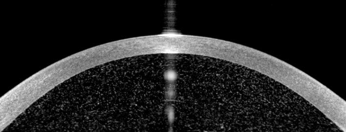
 Deep anterior segment OCT imaging
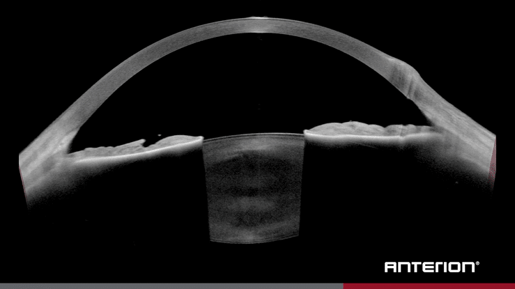
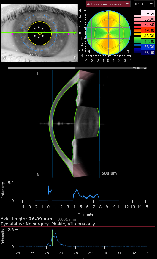
High-quality anterior segment OCT imaging allows for the visualization and assessment of the entire anterior chamber and lens. Combining longer wavelength swept-source OCT with eye tracking and image averaging (as on ANTERION*) provides rapid and repeatable high-resolution imaging of deep structures such as the ciliary muscle and the lens. This imaging approach may yield insights into muscle contraction and the dynamics of accommodation.
Related Publications
- Assessing accommodative presbyopic biometric changes of the entire anterior segment using single swept-source OCT image acquisitions (X. Xie, W. Sultan, G. Corradetti et al., 2021)
- Age- and refraction-related changes in anterior segment anatomical structures measured by swept-source anterior segment OCT (X. Xie, G. Corradetti, A. Song et al., 2020)
*Not available in all countries
 Epithelial thickness evaluation with high-resolution AS-OCT
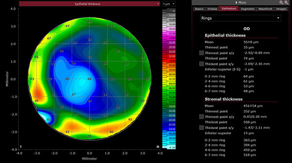
Evaluating epithelial thickness with a high-resolution anterior segment OCT provides the advantages of a non-contact method and simultaneous calculation of multiple corneal parameters. Thickness maps of the corneal epithelium and residual stroma may provide further insights for refractive surgery planning and treatment control, ectasia screening, and ocular surface disease.
Related Publications
- Epithelial Thickness Mapping in Keratoconic Corneas: Repeatability and Agreement Between CSO MS-39 Heidelberg Anterion and Optovue Avanti OCT Devices ( Y.Feng, D. Reinstein, T.Nitter et al,. 2023)
- Heidelberg Anterion Swept-Source OCT Corneal Epithelial Thickness Mapping: Repeatability and Agreement With Optovue Avanti (Y. Feng, D. Z. Reinstein, T. Nitter et al., 2022)
 Innovations in ectasia screening and monitoring
The early detection of ectatic change in the cornea can have significant impact on treatment planning and outcomes. The Screening Corneal Objective Risk of Ectasia (SCORE) Analyzer, developed by Dr. Gatinel and Dr. Saad (Paris, France), is an algorithm designed to support clinical decision-making. SCORE uses 12 discriminant indices to classify corneas according to the degree of similarity with those that are likely to progress to ectasia. The measurements are combined in a linear discriminant function, and all relevant metrics are multiplied by a coefficient to discriminate forme fruste keratoconus from normal corneas with a high level of sensitivity and specificity. SCORE is optimized for cornea data acquired with high-resolution swept-source OCT (ANTERION*) and aims to provide a modern method for screening and monitoring of keratoconus and other ectatic diseases.
Related Publications
- Discrimination between Keratoconus, Forme Fruste Keratoconus and Normal Eyes Using a Novel OCT-Based Tomographer (A. Saad, D. Gatinel, 2023)
- Diagnostic Validation of the Screening Corneal Objective Risk of Ectasia Analyzer Evaluated by Swept Source Optical Coherence Tomography for Keratoconus in an Asian Population (K. Kim, K. Koh, S. Lee et al., 2023)
- Ectasia Detection by Anterior Segment Optical Coherence Tomography in Scheimpflug Tomographically Normal Keratoconus Fellow Eyes (T. Naujokaitis, V. Augustin, H. Son et al. 2024)
- Automated Discrimination between Keratoconus, Forme Fruste Keratoconus and Normal Eyes Using a Novel OCT Based Tomographer (A. Saad, D. Gatinel, 2022)
*Not available in all countries
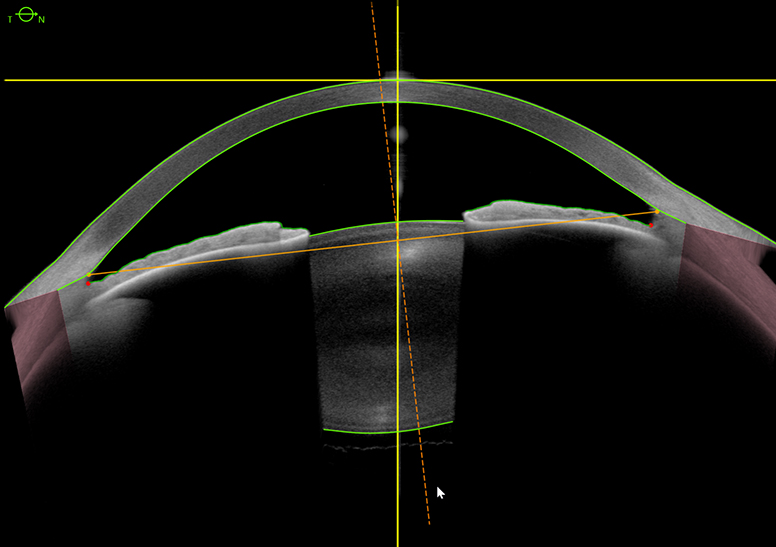
 Assessment of Anterior Chamber Inflammation with high-resolution Swept-Source AS-OCT
Anterior segment optical coherence tomography (AS-OCT) offers valuable insights into the quantification of anterior chamber inflammation in conditions such as uveitis and iritis. By providing high-resolution cross-sectional images of the anterior segment structures, AS-OCT enables precise measurement of parameters such as anterior chamber cell grading, corneal thickness, and flare intensity. This non-invasive imaging modality allows for objective evaluation of inflammation severity, guiding treatment decisions and monitoring disease progression. AS-OCT’s ability to visualize subtle changes in anterior segment morphology enhances diagnostic accuracy and facilitates timely intervention, ultimately improving patient outcomes in the management of anterior chamber inflammation.
Related Publications
- Establishing the Normative Data Set Necessary for Imaging-Based Childhood Uveitis Surveillance: A Cross-Sectional Study (A. Solebo, A. Bellchambers, S. Kellett et al., 2024)
- Anterior Segment Optical Coherence Tomography (AS-OCT) Visualization of Anterior Vitritis (F. Zicarelli, G. Staurenghi, A. Invernizzi., 2023)
 Keratometry Measurements and Dry Eye Disease
The current state of research concerning dry eye disease (DED) and its impact on the variability of keratometry measurements, and consequently, intraocular lens (IOL) power, presents conflicting findings. This highlights the need to further explore the influence of DED on ocular biometric measurements crucial for calculating IOL power.
Related Publications
 Artificial Intelligence in Angle Closure Disease
Growing evidence supports the clinical usefulness of anterior segment optical coherence tomography (AS-OCT) for assessing anterior segment biometrics, particularly scleral spur-based measurements. Quantitative AS-OCT methods could complement gonioscopy, the current standard for assessing the anterior chamber angle (ACA), which is subjective and variably reproducible. The expertise-dependent and time-consuming process of manual marking of scleral spurs however hinders widespread AS-OCT implementation in routine clinical practice. Leveraging deep learning algorithms (as on ANTERION*) enables accurate scleral spur detection and biometric analysis, offering a solution to these challenges and facilitating broader implementation of AS-OCT.
Related Publications

Please note: If not indicated differently, the technologies mentioned are not for sale and are not a reflection of our commercial product releases or planned product developments.


