You are currently on the Cocos (Keeling) Islands version.
Looks like you are in United States.
Switch to local content?














Analyze the Cornea at the Cellular Level
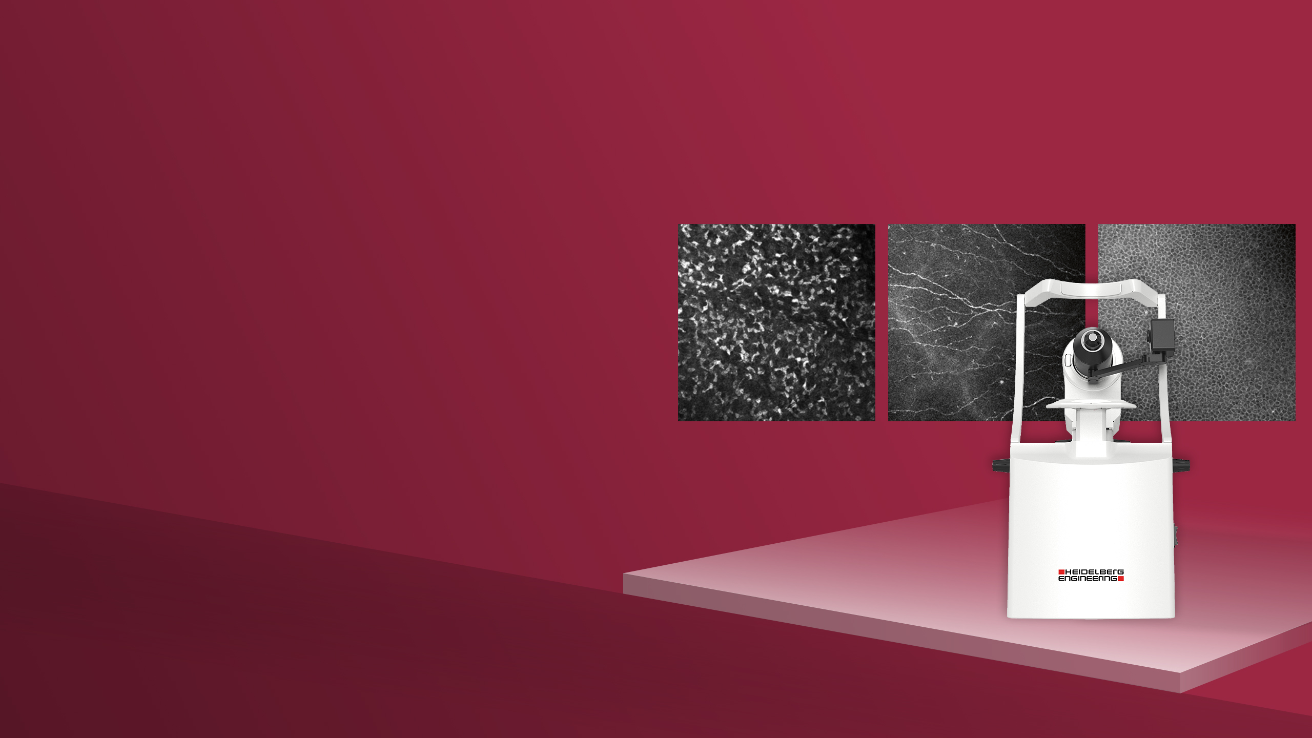
Heidelberg RCM allows you to acquire unique en face images of corneal cells and microstructures non-invasively. Examine keratocyte subpopulations, visualize the fine details of the subbasal nerve plexus and assess endothelial cell morphology.
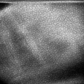
Epithelium
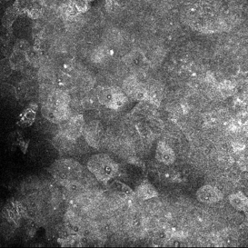
Visualize epithelial cells and assess their morphology at a selected imaging depth.
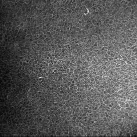
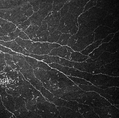
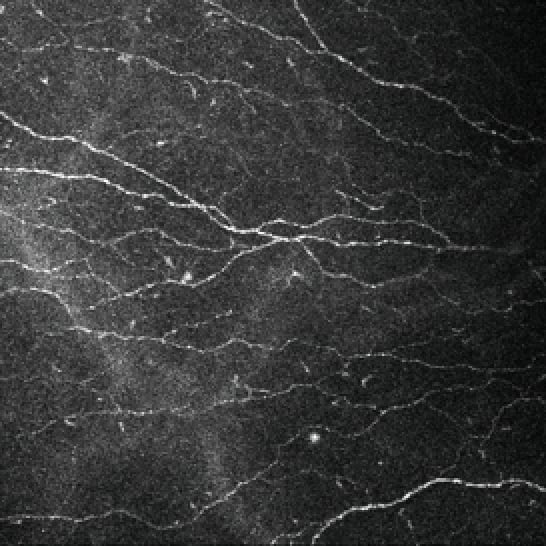
Subbasaler Nervenplexus
Untersuchen Sie das Netzwerk der Hornhautnerven und prüfen Sie die Nervenmorphologie im Detail.
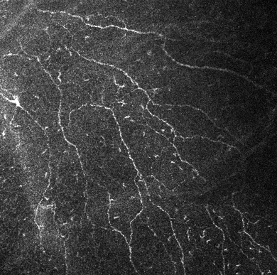
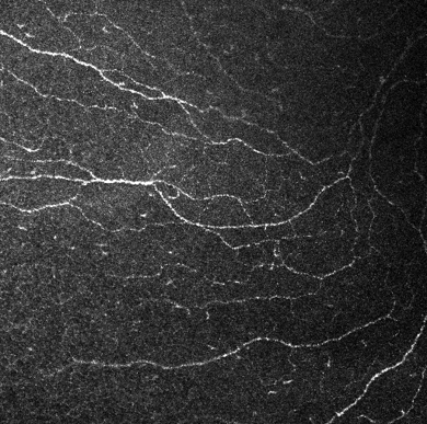
Stroma
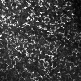
Explore stromal microstructure at different imaging depths and visualize the morphology of structures including keratocytes.
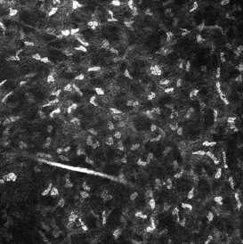
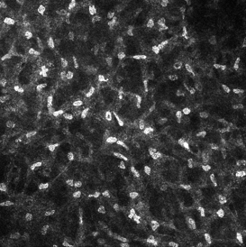
Endothelium
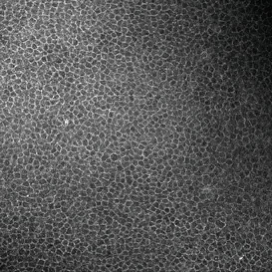
Visualize the corneal endothelium and assess cellular morphology.
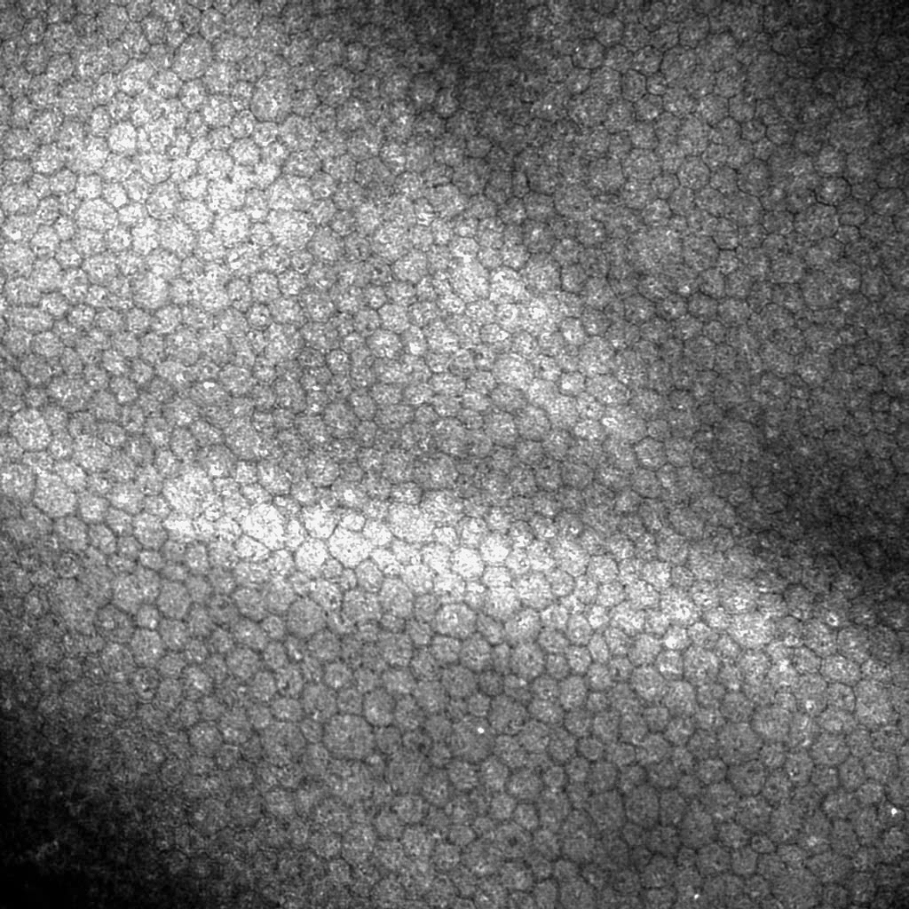
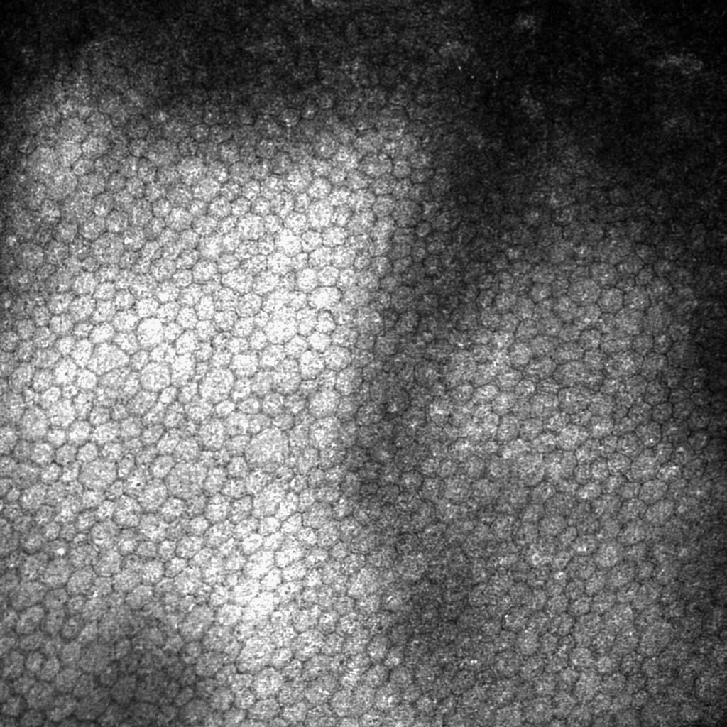
Image courtesy: Gaush Meditech
Image courtesy: Gaush Meditech