Side-by-side evaluations show MultiColor provides better visualization of certain pathologies
Heidelberg, Germany – Multiple international studies evaluating the sensitivity of SPECTRALIS MultiColor confocal scanning laser ophthalmoscopy versus color fundus photography were presented at the Association for Vision and Research in Ophthalmology (ARVO) 2018 conference in Honolulu, Hawaii. The studies presented at ARVO reported better sensitivity and visualization of a number of pathologies using the SPECTRALIS MultiColor Module in comparison to color fundus photography.
The SPECTRALIS MultiColor Module differs from color fundus photography in that it uses three laser wavelengths simultaneously and a 5μm/pixel resolution to provide diagnostic images that show distinct structures at different depths within the retina and clearer images than conventional color fundus photography.
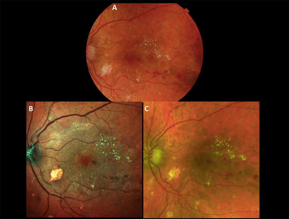
Amit Meshi, UCSD, San Diego, California, USA, et al.1 compared retinal pathology visualization and detection between the SPECTRALIS MultiColor Module, Optos P200 and Topcon TRC-50DX. The authors concluded that while the overall detection rate of retinal pathology was similar across all three devices, a larger area of macular involvement and significantly better epiretinal membrane (ERM) visualization were noted on the SPECTRALIS MultiColor images. A comparison of the three technologies is shown in figure 1.
A study by Ryoh Funatsu, Kagoshima University, Kagoshima-shi, Japan, et al.2 was in agreement with the findings by Amit Meshi et al. and concluded that “evaluation of ERM by MultiColor is more sensitive than color fundus photography” and in addition reported that “MultiColor might be a possible marker for metamorphopsia in ERM patients.”
For the identification of diabetic maculopathy, Obaid Kousha, Ninewells Hospital, London, et al.3 showed that MultiColor combined with SD-OCT provides “effective monitoring” of diabetic maculopathy and that “5 eyes with exudates and severe macular edema requiring urgent intervention were missed by color fundus photography but not by MultiColor.” The benefit of multimodal imaging was highlighted by their finding that “MultiColor, when complemented by SD-OCT, didn’t miss any clinically significant macular edema.”
Hiroto Terasaki, Kagoshima University, Kagoshima-shi, Japan, et al.4 compared fundus images obtained by MultiColor to those obtained using color fundus photography in detecting nerve fiber layer defects. They found that “detection of abnormal findings in the nerve fiber layer by MultiColor was more sensitive than by color fundus photography.”
Find out more about the MultiColor Module for SPECTRALIS at www.spectralis-multicolor.com.
- Amit Meshi, UCSD, San Diego, California, USA, et al. Comparison of retinal pathology visualization in multi-spectral scanning laser imaging. Abstract Number: 1501 – C0342, ARVO 2018.
- Ryoh Funatsu, Kagoshima University, Kagoshima-shi, Japan, et al. Comparison of color fundoscopy and SPECTRALIS MultiColor on detection of epiretinal membrane and relationship between ERM findings and metamorphopsia. Posterboard Number: C0111, ARVO 2018.
- Obaid Kousha, Ninewells Hospital, London, et al. Diabetic maculopathy: Multicolor and SD-OCT versus fundus photography. Posterboard Number: C0111, ARVO 2018.
- Hiroto Terasaki, Kagoshima University, Kagoshima-shi, Japan, et al. Diabetic maculopathy: Multicolor and SD-OCT versus fundus photography. Presentation Number: 3444, ARVO 2018