A Sudden Change in the Visual Fields
In this case study, Dr. James Fanelli describes how a sudden, dramatic change in a glaucoma patient’s visual field initially raised concerns about disease progression but was ultimately traced to upper eyelid dermatochalasis obstructing the patient’s vision—an observation made through a physical exam and conversation about patient habits. Heidelberg Engineering imaging technology allowed the clinician/the doctor to further confirm structural stability, demonstrating the importance of combining advanced imaging with clinical observation in glaucoma management.
Read the full Optometry Times article here
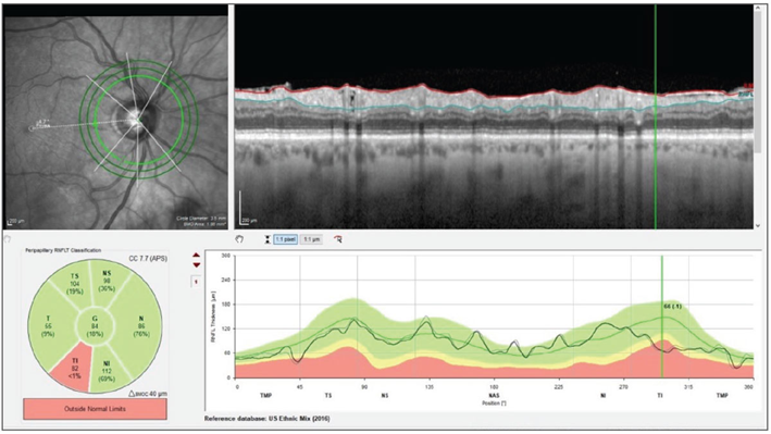
Image courtesy: James L. Fanelli, OD


