You are currently on the Isle of Man version.
Looks like you are in United States.
Switch to local content?












Heidelberg Engineering Announces Commercial Availability for new SPECTRALIS® Software Release, Advancing Speed and Diagnostic Confidence
Heidelberg Engineering announced the commercial availability of its CE and FDA cleared latest SPECTRALIS® Software Version.
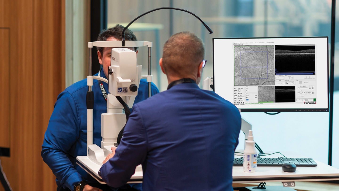
HEIDELBERG, GERMANY – 31.08.2026 – Heidelberg Engineering, a global leader in ophthalmic imaging and healthcare data solutions, today announced the commercial availability of its CE and FDA cleared latest SPECTRALIS® Software Version. This release is designed to deliver speed, workflow flexibility, and diagnostic depth to multimodal retinal imaging.
Under the theme ‘Faster, Reliable, Patient-centered’, the significant enhancements of SPECTRALIS® introduce 250 kHz scan speed for OCT angiography (OCTA) combined with enhanced TruTrack Active Eye Tracking. New multimodal imaging capabilities, including the Green Autofluorescence Module, designed to further expand clinical insight while maintaining the trusted Heidelberg image quality clinicians rely on for consistent, reliable data.
“This new SPECTRALIS® software release reflects our continued commitment to advancing ophthalmic imaging,” said Holger Ruchatz, Head of Product Management at Heidelberg Engineering. “By bringing together speed, precision, and multimodal imaging, it is designed to support clinicians in visualizing and assessing retinal disease.”
SPECTRALIS® with SHIFT Technology
The latest release on SPECTRALIS® with SHIFT Technology introduces 250 kHz scan speed for OCT angiography imaging, complementing the existing 85 kHz, 125 kHz, and 20 kHz scan speeds for OCT and OCTA imaging. This multi-speed approach allows clinicians to remain flexible and tailor imaging to workflow demands and clinical needs, balancing acquisition time and image detail for each patient.
The addition of Dense ART, with ART values up to 100 for OCT angiography imaging, allows for image averaging over a small area for the visualization of specific regions of interest for more detailed interrogation of both structure and flow in vascular pathologies. With this feature, clinicians can image vasculature with confidence and support timely treatment decisions.
Green Autofluorescence Imaging
The Green Autofluorescence Module improves visualization of the macula, enabling identification of key features such as foveal sparing in macular atrophy. This clarity allows for confident diagnosis, patient selection, and disease monitoring, while compatibility with RegionFinder enables quantification of atrophy for clinical assessment and longitudinal analysis. The combination of Green Autofluorescence and BluePeak blue autofluorescence imaging provides a comprehensive view of retinal structure and metabolic changes, offering deeper clinical insight.
Additional features further support clinical workflow, patient comfort, and advanced imaging capabilities. Visit our Product Page or contact us directly for more information. Visit us at ESCRS and EURETINA 2026 to experience the new SPECTRALIS® Software Version in person.
DISCLAIMER: This content may contain product information differing from the current approval status in your country. Please refer to your country-specific website for relevant information about your market.