BluePeak Module
Blue Autofluorescence
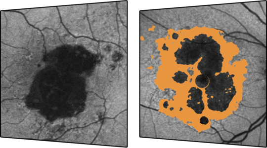
BluePeak autofluorescence is a non-invasive scanning laser fundus imaging modality that reveals metabolic stress in the retina using lipofuscin as an indicator. BluePeak images can reveal RPE and photoreceptor cell malfunctions, offering diagnostic insights into retinal conditions such as AMD and hereditary diseases.
By combining the segmentation of retinal layers in OCT with the metabolic mapping of BluePeak autofluorescence, morphologic alterations associated with functional change can be seen more clearly.